
Lab 7: Muscular System II – Forelimb and Axial Muscles
Muscles of the forelimb, trunk, and head; Muscle groups
Learning Objectives
When you are prepared for the Test on Week 6-7 Learning Objectives in Week 8*, you will be able to:
- Identify components of a muscle cell and the neuromuscular junction.
- Identify connective tissues of muscles.
- Identify significant muscles on both human models and cats (listed in the lab activity).
- For the hamstrings, quadriceps and rotator cuff muscle groups: identify the muscles within them and the bony features that act as their attachment sites.
*Muscle dissections will span weeks 6-7. There will be no test in Week 7. In week 8, there will be a test covering all of the above learning objectives.
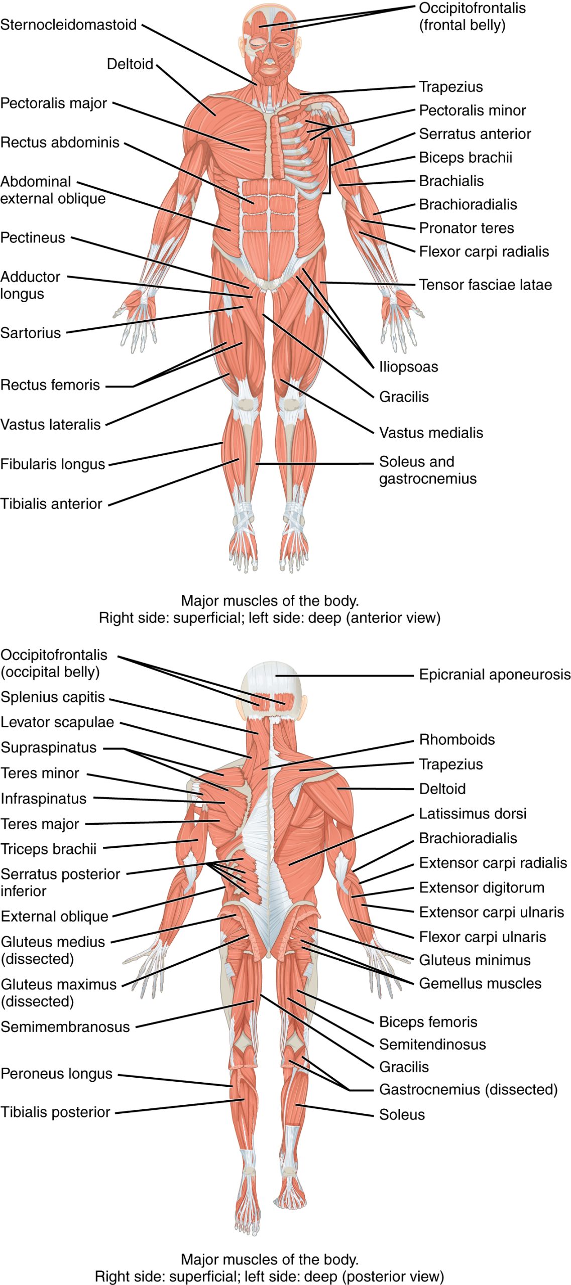
In last week’s lab, we looked at muscles of the hindlimb. In lab this week, we will look at the skeletal muscles in the forelimb, as well as in the axial region. By the end of these two weeks, you should have a broad understanding of the muscular system and be able to identify the major muscles in the body (Figure 7.1).
Muscles of the Forelimb
Muscles of the shoulder and upper limb can be divided into four groups: muscles that stabilize and position the shoulder girdle, muscles that move the arm, muscles that move the forearm, and muscles that move the wrists, hands, and fingers. The shoulder girdle consists of the lateral ends of the clavicle and scapula, along with the proximal end of the humerus, and the muscles covering these three bones to stabilize the shoulder joint. The girdle creates a base from which the head of the humerus, in its ball-and-socket joint with the glenoid fossa of the scapula, can move the arm in multiple directions.
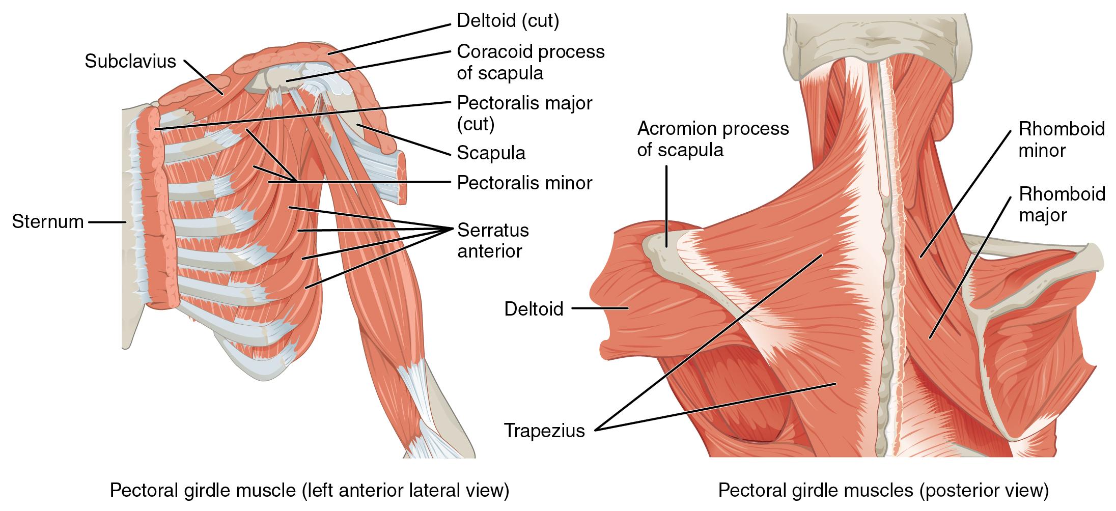
Muscles That Position the Pectoral Girdle
Muscles that position the pectoral girdle are located either on the anterior thorax or on the posterior thorax (Figure 7.2 and Table 7.1). The anterior muscles include the subclavius, pectoralis minor, and serratus anterior. The posterior muscles include the trapezius, rhomboid major, and rhomboid minor. When the rhomboids are contracted, your scapula moves medially, which can pull the shoulder and upper limb posteriorly.
| Position in the thorax | Movement | Target | Target motion direction | Prime mover | Origin | Insertion |
|---|---|---|---|---|---|---|
| Anterior thorax | Stabilizes clavicle during movement by depressing it | Clavicle | Depression | Subclavius | First rib | Inferior surface of clavicle |
| Anterior thorax | Rotates shoulder anteriorly (throwing motion); assists with inhalation | Scapula; ribs | Scapula: depresses; ribs: elevates | Pectoralis minor | Anterior surfaces of certain ribs (2–4 or 3–5) | Coracoid process of scapula |
| Anterior thorax | Moves arm from side of body to front of body; assists with inhalation | Scapula; ribs | Scapula: protracts; ribs: elevates | Serratus anterior | Muscle slips from certain ribs (1–8 or 1–9) | Anterior surface of vertebral border of scapula |
| Posterior thorax | Elevates shoulders (shrugging); pulls shoulder blades together; tilts head backwards | Scapula; cervical spine | Scapula: rotests inferiorly, retracts, elevates, and depresses; spine: extends | Trapezius | Skull; vertebral column | Acromion and spine of scapula; clavicle |
| Posterior thorax | Stabilizes scapula during pectoral girdle movement | Scapula | Retracts; rotates inferiorly | Rhomboid major | Thoracic vertebrae (T2–T5) | Medial border of scapula |
| Posterior thorax | Stabilizes scapula during pectoral girdle movement | Scapula | Retracts; rotates inferiorly | Rhomboid minor | Cervical and thoracic vertebrae (C7 and T1) | Medial border of scapula |
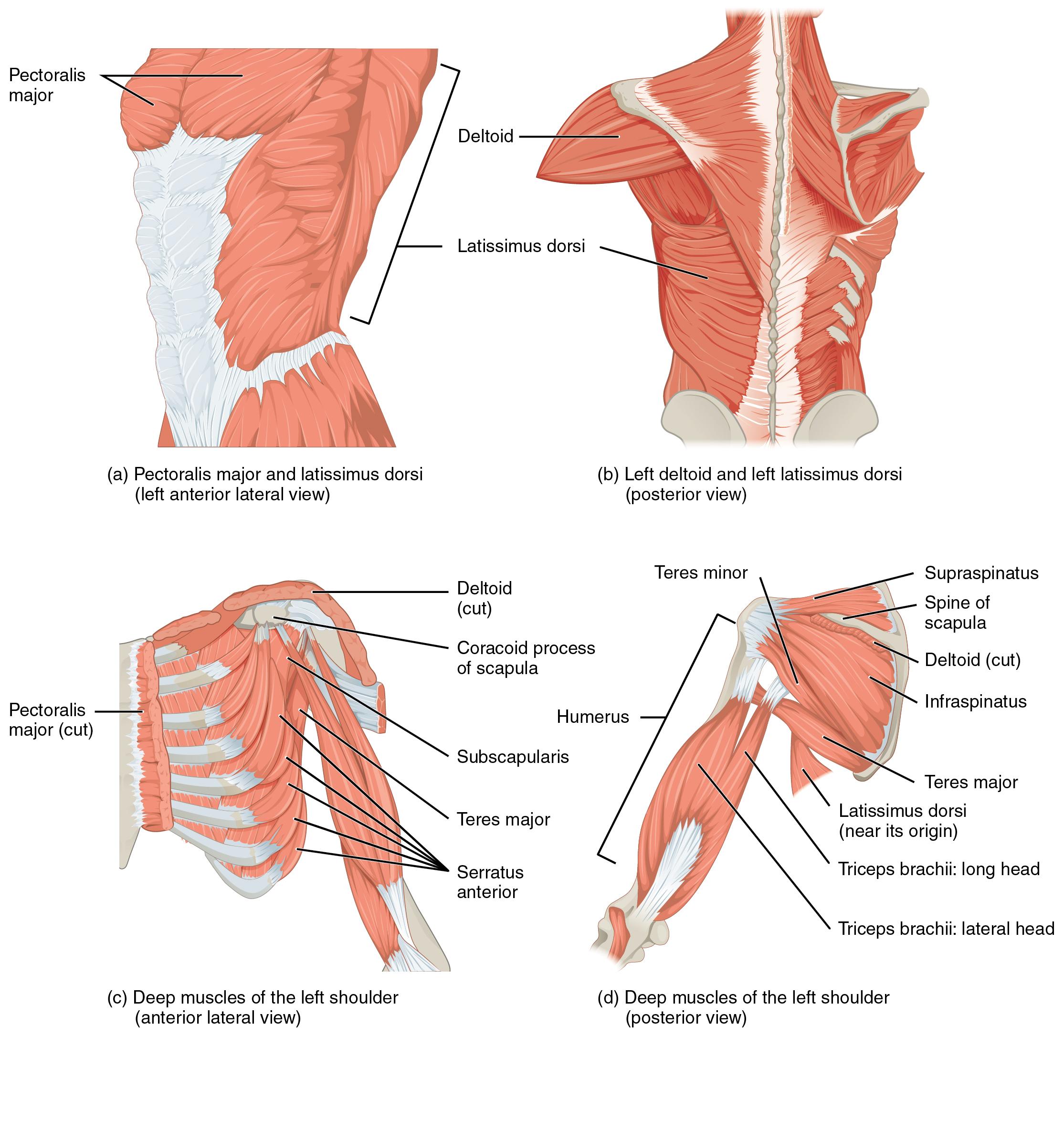
Muscles That Move the Humerus
Similar to the muscles that position the pectoral girdle, muscles that cross the shoulder joint and move the humerus bone of the arm include both axial and scapular muscles (Figure 7.3 and Table 7.2). The two axial muscles are the pectoralis major and the latissimus dorsi. The pectoralis major is thick and fan-shaped, covering much of the superior portion of the anterior thorax. The broad, triangular latissimus dorsi is located on the inferior part of the back, where it inserts into a thick connective tissue sheath called an aponeurosis.
| Group | Movement | Target | Target motion direction | Prime mover |
Origin | Insertion |
|---|---|---|---|---|---|---|
| Axial Muscles | Brings elbows together; moves elbow up (as during an uppercut punch) | Humerus | Flexion; adduction; medial rotation | Pectoralis major | Clavicle; sternum; cartilage of certain ribs (1-6 or 1-7); aponeurosis of external oblique muscle | Greater tubercle of humerus |
| Moves elbow back (as in elbowing someone standing behind you); spreads elbows apart | Humerus; scapula | Humerus; extension, adduction, and medial rotation; scapula: depression | Latissimus dorsi | Thoracic vertebrae (T7-T12); lumbar vertebrae; lower ribs (9-12); iliac crest | Intertubercular sulcus of humerus | |
| Rotator Cuff
(scapular muscles) |
Assists pectoralis major in bringing elbows together ans stabilizes shoulder joint during movement of the pectoral girdle | Humerus | Medial rotation | Subscapularis | Subscapular fossa of scapula | Lesser tubercle of humerus |
| Rotates elbow outwards, as during a tennis swing | Humerus | Abduction | Supraspinatus | Supraspinous foss of scapula | Greater tubercle of humerus | |
| Rotates elbow outwards, as during a tennis swing | Humerus | Extension; adduction | Infraspinatus | Infraspinous fossa of scapula | Greater tubercle of humerus | |
| Assists infraspinatus in rotating elbow outwards | Humerus | Extension; adduction | Teres minor | Lateral border of dorsal scapular surface | Greater tubercle of humerus | |
| Other scapular muscles | Assists infraspinatus in rotating elbow outwards | Humerus | Extension; adduction | Teres major | Posterior surface of scapula | Intertubercular sulcus of humerus |
| Lifts arms at shoulder | Humerus | Abduction; flexion; extension; medial and lateral rotation | Deltoid | Trapezius; clavicle; acromion; spine of scapula | Deltoid tuberosity of humerus | |
| Moves elbow up and across body, as when putting hand on chest | Humerus | Flexion; adduction | Coracobrachialis | Coracoid process of scapula | Medial surface of humerus shaft |
The rest of the shoulder muscles originate on the scapula. The anatomical and ligamental structure of the shoulder joint and the arrangements of the muscles covering it, allows the arm to carry out different types of movements. The deltoid, the thick muscle that creates the rounded lines of the shoulder is the major abductor of the arm, but it also facilitates flexing and medial rotation, as well as extension and lateral rotation. The subscapularis originates on the anterior scapula and medially rotates the arm. Named for their locations, the supraspinatus (superior to the spine of the scapula) and the infraspinatus (inferior to the spine of the scapula) abduct the arm, and laterally rotate the arm, respectively. The thick and flat teres major is inferior to the teres minor and extends the arm, and assists in adduction and medial rotation of it. The long teres minor laterally rotates and extends the arm. Finally, the coracobrachialis flexes and adducts the arm.
The tendons of the deep subscapularis, supraspinatus, infraspinatus, and teres minor connect the scapula to the humerus, forming the rotator cuff (musculotendinous cuff), the circle of tendons around the shoulder joint. When baseball pitchers undergo shoulder surgery it is usually on the rotator cuff, which becomes pinched and inflamed, and may tear away from the bone due to the repetitive motion of bring the arm overhead to throw a fast pitch.
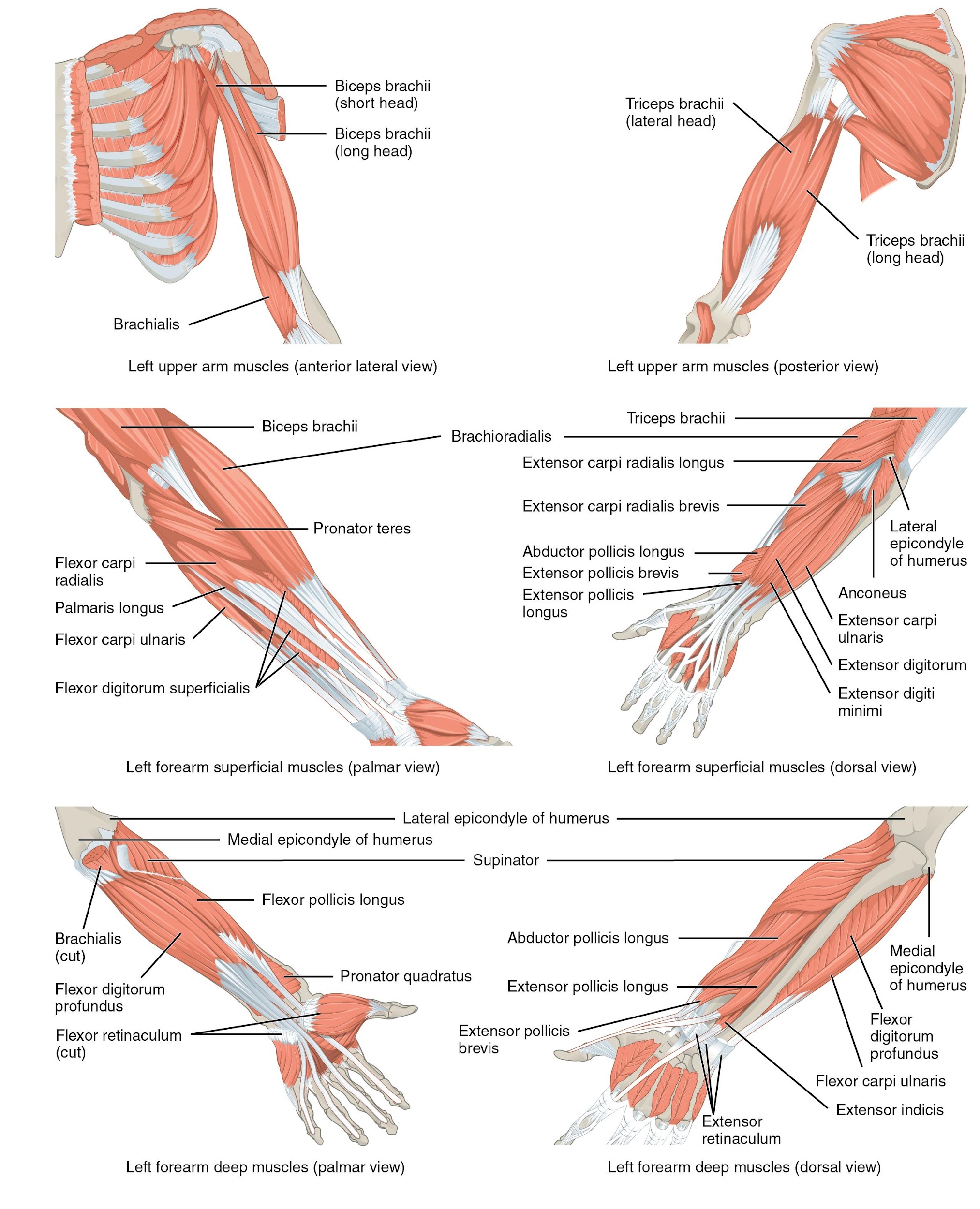
Muscles That Move the Forearm
The forearm, made of the radius and ulna bones, has four main types of action at the hinge of the elbow joint: flexion, extension, pronation, and supination. The elbow flexors include the biceps brachii, brachialis, and brachioradialis. The elbow extensor is the triceps brachii. The forearm pronators are the pronator teres and the pronator quadratus, and the supinator is the only one that supinates the forearm (turns the palm of the hand anteriorly).
The biceps brachii, brachialis, and brachioradialis flex the elbow. The two-headed biceps brachii crosses the shoulder and elbow joints to flex the elbow, also taking part in supinating the forearm at the radioulnar joints and flexing the arm at the shoulder joint. Deep to the biceps brachii, the brachialis provides additional power in flexing the elbow. Finally, the brachioradialis can flex the elbow quickly or help lift a load slowly. These muscles and their associated blood vessels and nerves form the anterior compartment of the arm (anterior flexor compartment of the arm) (Figure 7.4 and Table 7.3).
| Movement | Target | Target Motion Direction |
Prime Mover
|
Origin
|
Insertion
|
|---|---|---|---|---|---|
| Anterior Muscles (Flexion) | |||||
| Performs a bicep curl; also allows palm of hand to point toward body while flexing |
Forearm | Flexion; supination | Biceps brachii | Coracoid process; tubercle above glenoid cavity | Radial tuberosity |
| Performs a bicep curl | Forearm | Flexion | Brachialis | Front of distal humerus | Coronoid process of ulna |
| Assists and stabilizes elbow during bicep curl motion | Forearm | Flexion | Brachioradialis | Lateral supracondylar ridge at distal end of humerus | Base of styloid process of radius |
| Posterior Muscles (Extension) | |||||
| Assists in extending forearm; also allows forearm to extend away from the body | Forearm | Extension; abduction | Anconeus | Lateral epicondyle of humerus |
Lateral aspect of olecranon process of ulna |
| Extends forearm, as during a punch | Forearm | Extension | Triceps brachii | Infraglenoid tubercle of scapula ; posterior shaft of humerus ; posterior humeral shaft distal to the radial groove |
Olecranon process of ulna |
| Anterior Muscles (Pronation) | |||||
| Assists in turning hand palm down |
Forearm | Pronation | Pronator quadratus | Distal portion of anterior ulnar shaft |
Distal surface of anterior radius |
| Turns hand palm down |
Forearm | Pronation | Pronator teres | Medial epicondyle of humerus ; coronoid process of ulna |
Lateral radius |
| Posterior Muscles (Supination) | |||||
| Turns hand palm up |
Forearm | Supination | Supinator | Lateral epicondyle of humerus; proximal ulna | Proximal end of radius |
Muscles That Move the Wrist, Hand, and Fingers
Wrist, hand, and finger movements are facilitated by two groups of muscles. The forearm is the origin of the extrinsic muscles of the hand. The palm is the origin of the intrinsic muscles of the hand.
The muscles in the anterior compartment of the forearm (anterior flexor compartment of the forearm) originate on the humerus and insert into different parts of the hand. These make up the bulk of the forearm. From lateral to medial, the superficial anterior compartment of the forearm includes the flexor carpi radialis, palmaris longus, flexor carpi ulnaris, and flexor digitorum superficialis. The flexor digitorum superficialis flexes the hand as well as the digits at the knuckles, which allows for rapid finger movements, as in typing or playing a musical instrument (see Table 7.4). However, poor ergonomics can irritate the tendons of these muscles as they slide back and forth with the carpal tunnel of the anterior wrist and pinch the median nerve, which also travels through the tunnel, causing Carpal Tunnel Syndrome. The deep anterior compartment produces wrist and digit flexion, bending the fingers to make a fist. These are the flexor pollicis longus and the flexor digitorum profundus.
The muscles in the superficial posterior compartment of the forearm (superficial posterior extensor compartment of the forearm) originate on the humerus. These are the extensor radialis longus, extensor carpi radialis brevis, extensor digitorum, extensor digiti minimi, and the extensor carpi ulnaris.
The muscles of the deep posterior compartment of the forearm (deep posterior extensor compartment of the forearm) originate on the radius and ulna. These include the abductor pollicis longus, extensor pollicis brevis, extensor pollicis longus, and extensor indicis.
The tendons of the forearm muscles attach to the wrist and extend into the hand. Fibrous bands called retinacula sheath the tendons at the wrist. The flexor retinaculum extends over the palmar surface of the hand while the extensor retinaculum extends over the dorsal surface of the hand.
| Movement | Target |
Target motion direction |
Prime mover | Origin | Insertion |
|---|---|---|---|---|---|
| Superficial anterior compartment of forearm | |||||
| Wrist flexion and abduction | Wrist; hand | Flexion; abduction | Flexor carpi radialis | Medial epicondyle of humerus | Base of 2nd and 3rd metacarpals |
| Wrist flexion and adduction | Wrist | Flexion | Flexor carpi ulnaris | Medial epicondyle of humerus | Pisiform, hamate, base of 5th metacarpal |
| Wrist flexion; finger flexion digits 2-5 | Wrist; hand | Flexion; adduction | Flexor digitorum superficialis | Medial epicondyle of humerus; anterior shaft of radius | Middle phalanges of digits 2-5 |
| Wrist flexion | Wrist; fingers 2-5 | Flexion | Palmaris longus | Medial epicondyle of humerus | Palmar aponeurosis |
| Deep anterior compartment of forearm | |||||
| Wrist flexion; finger flexion digits 2-5 | Thumb | Flexion | Flexor digitorum profundus | Coronoid process; anteromedial surface of ulna | Distal phalanges of digits 2-5 |
| Thumb flexion | Wrist; fingers | Flexion | Flexor pollicis longus | Anterior surface of radius | Distal phalanx of thumb |
| Superficial posterior compartment of forearm | |||||
| Wrist extension and abduction | Wrist | Extension; abduction | Extensor carpi radialis longus | Lateral supracondylar ridge of humerus | Base of 2nd metacarpal |
| Wrist extension and abduction | Wrist | Extension; abduction | Extensor carpi radialis brevis | Lateral epicondyle of humerus | Base of 3rd metacarpal |
| Wrist extension and adduction | Wrist; fingers | Extension; abduction | Extensor carpi ulnaris | Lateral epicondyle of humerus | Base of 5th metacarpal |
| Wrist extension; finger extension | Little fingers | Extension | Extensor digitorum | Lateral epicondyle of humerus | Extensor expansions; distal phalanges of digits 2-5 |
| Finger extension of digit 5 | Wrist | Extension; adduction | Extensor digiti minimi | Lateral epicondyle of humerus | Extensor expansions; distal phalanx of digit 5 |
| Deep posterior compartment of forearm | |||||
| Thumb abduction and extensionWrist abduction | Wrist; thumb | Thumb: abduction, extension; wrist: abduction | Abductor pollicis longus | Posterior surface of radius and ulna | Base of first metacarpal; trapezium |
| Thumb extension | Thumb | Extension | Extensor pollicis longus | Posterior shaft of radius and ulna | Base of distal phalanx of thumb |
| Thumb extension | Thumb | Extension | Extensor pollicis brevis | Posterior shaft of radius and ulna | Base of proximal phalanx of thumb |
| Finger extension of digit 2 | Wrist; index finger | Extension | Extensor indicis | Posterior surface of radius and ulna | Tendon of extensor digitorum of index finger |
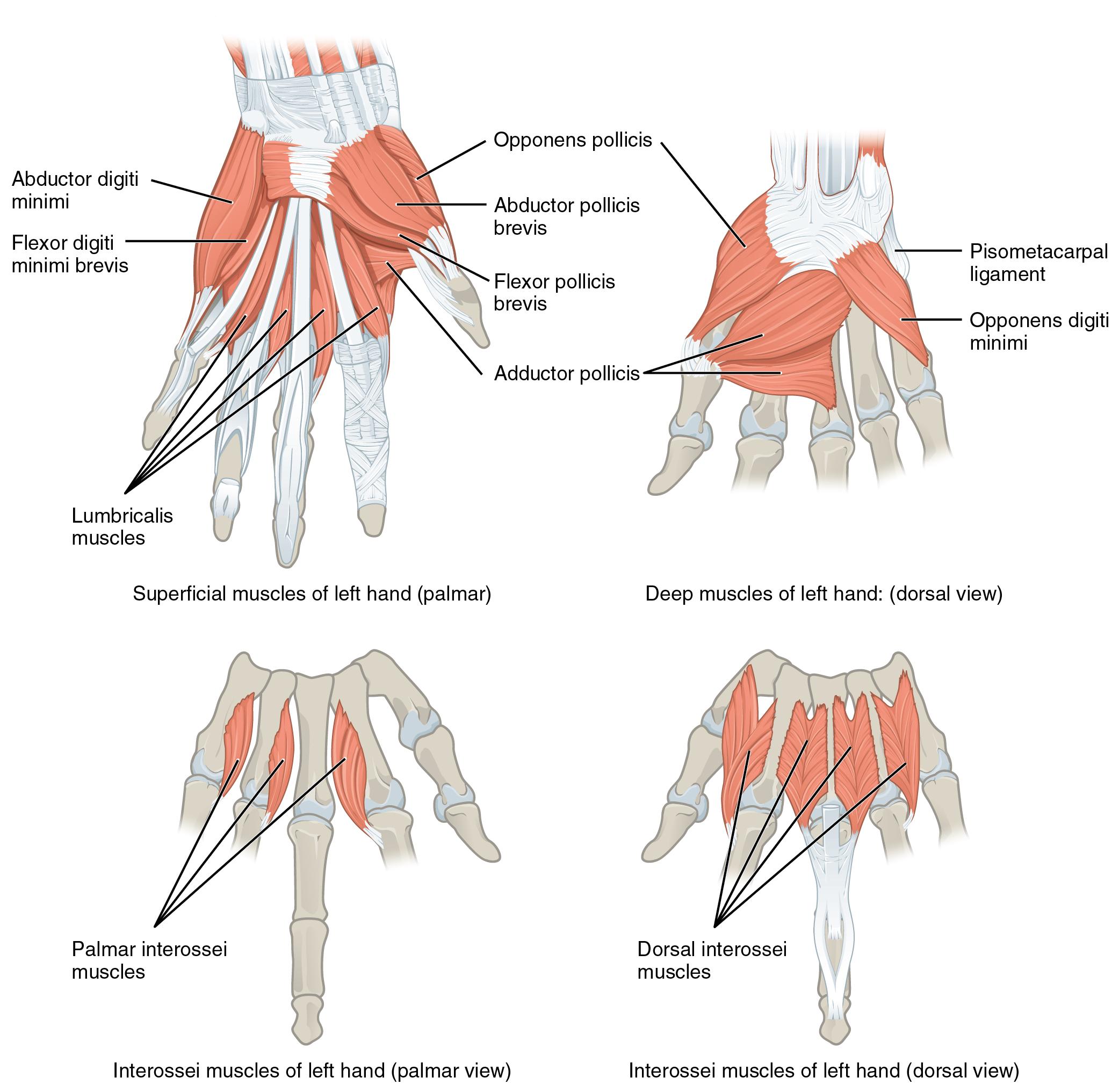
Intrinsic Muscles of the Hand
The intrinsic muscles of the hand both originate and insert within it (Figure 7.5). These muscles allow your fingers to also make precise movements for actions, such as typing or writing. These muscles are divided into three groups. The thenar muscles are on the radial aspect of the palm. The hypothenar muscles are on the medial aspect of the palm, and the intermediate muscles are midpalmar.
The thenar muscles include the abductor pollicis brevis, opponens pollicis, flexor pollicis brevis, and the adductor pollicis. These muscles form the thenar eminence, the rounded contour of the base of the thumb, and all act on the thumb. The movements of the thumb play an integral role in most precise movements of the hand.
The hypothenar muscles include the abductor digiti minimi, flexor digiti minimi brevis, and the opponens digiti minimi. These muscles form the hypothenar eminence, the rounded contour of the little finger, and as such, they all act on the little finger. Finally, the intermediate muscles act on all the fingers and include the lumbrical, the palmar interossei, and the dorsal interossei.
Axial Muscles
The skeletal muscles are divided into axial (muscles of the trunk and head) and appendicular (muscles of the arms and legs) categories. The axial muscles are grouped based on location, function, or both. Some of the axial muscles may seem to blur the boundaries because they cross over to the appendicular skeleton. The first grouping of the axial muscles you will review includes the muscles of the head and neck, then you will review the muscles of the vertebral column, and finally you will review the oblique and rectus muscles.
Muscles That Create Facial Expression
The origins of the muscles of facial expression are on the surface of the skull (remember, the origin of a muscle does not move). The insertions of these muscles have fibers intertwined with connective tissue and the dermis of the skin. Because the muscles insert in the skin rather than on bone, when they contract, the skin moves to create facial expression (Figure 7.6 and Table 7.5).
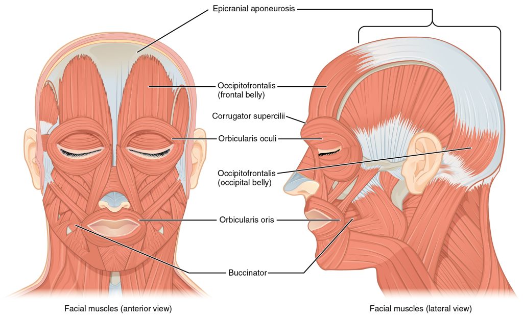
The orbicularis oris is a circular muscle that moves the lips, and the orbicularis oculi is a circular muscle that closes the eye. The occipitofrontalis muscle moves up the scalp and eyebrows. The muscle has a frontal belly and an occipital (near the occipital bone on the posterior part of the skull) belly. In other words, there is a muscle on the forehead (frontalis) and one on the back of the head (occipitalis), but there is no muscle across the top of the head. Instead, the two bellies are connected by a broad tendon called the epicranial aponeurosis, or galea aponeurosis (galea = “helmet”). The physicians originally studying human anatomy thought the skull looked like a helmet.
A large portion of the face is composed of the buccinator muscle, which compresses the cheek. This muscle allows you to whistle, blow, and suck; and it contributes to the action of chewing. There are several small facial muscles, one of which is the corrugator supercilii, which is the prime mover of the eyebrows. Place your finger on your eyebrows at the point of the bridge of the nose. Raise your eyebrows as if you were surprised and lower your eyebrows as if you were frowning. With these movements, you can feel the action of the corrugator supercilii.
| Movement | Target | Target Motion Direction | Primer Mover | Origin | Insertion |
|---|---|---|---|---|---|
| Brow | |||||
| Raising eyebrows (e.g. showing surprise) | Skin of scalp | Anterior | Occipitofrontalis (front belly) | Epicraneal aponeurosis | Underneath skin of forehead |
| Tensing and retracting scalp | Skin of scalp | Posterior | Occipitofrontalis (occipital belly) | Occipital bone; mastoid process (temporal bone) | Epicranial aponeurosis |
| Lowering eyebrows (e.g. scowling, frowning) | Skin underneath eyebrows | Inferior | Corrugator supercilii | Frontal bone | Skin underneath eyebrow |
| Nose | |||||
| Flaring nostrils | Nasal cartilage (pushes nostrils open when cartilage is compressed) | Inferior compression; posterior compression | Nasalis | Maxilla | Nasal bone |
| Mouth | |||||
| Raising upper lip | Upper lip | Elevation | Levator labii superioris | Maxilla | Underneath skin at corners of the mouth; prbicularis oris |
| Lowering lower lip | Lower lip | Depression | Depressor labii inferioris | Mandible | Underneath skin of lower lip |
| Opening mouth and sliding lower jaw left and right | Lower jaw | Depression, lateral | Depressor angulus oris | Mandible | Underneath skin at corners of mouth |
| Smiling | Corners of mouth | Lateral elevation | Zygomaticus major | Zygomatic bone | Underneath skin at corners of mouth (dimple area); orbicularis oris |
| Shaping of lips (during speech) | Lips | Multiple | Orbicularis oris | Tissue surrounding lips | Underneath skin at corners of mouth |
| Lateral movement of cheeks (e.g. sucking on a straw; also used to compress air in mouth while blowing) | Cheeks | Lateral | Buccinator | Maxilla, mandible; sphenoid bone (via pterygomandibular raphe) | Orbicularis oris |
| Pursing of lips by straightening them laterally | Corners of mouth | Lateral | Risorius | Fascia of parotid salivary gland | Underneath skin at corners of the mouth |
| Protrusion of lower lip (e.g. pouting expression) | Lower lip and skin of chin | Protraction | Mentalis | Mandible | Underneath skin of chin |
Muscles That Move the Lower Jaw
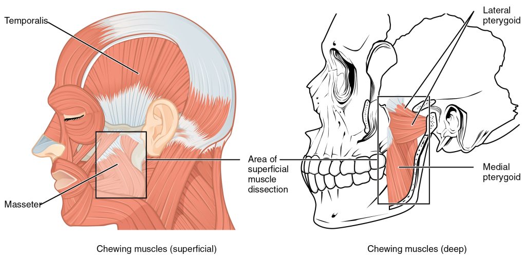
In anatomical terminology, chewing is called mastication. Muscles involved in chewing must be able to exert enough pressure to bite through and then chew food before it is swallowed (Figure 7.7). The masseter muscle is the main muscle used for chewing because it elevates the mandible (lower jaw) to close the mouth, and it is assisted by the temporalis muscle, which retracts the mandible. You can feel the temporalis move by putting your fingers to your temple as you chew. Although the masseter and temporalis are responsible for elevating and closing the jaw to break food into digestible pieces, the medial pterygoid and lateral pterygoid muscles provide assistance in chewing and moving food within the mouth.
Muscles of the Anterior Neck
The muscles of the anterior neck assist in deglutition (swallowing) and speech by controlling the positions of the larynx (voice box), and the hyoid bone, a horseshoe-shaped bone that functions as a solid foundation on which the tongue can move. The muscles of the neck are categorized according to their position relative to the hyoid bone (Figure 7.8). Suprahyoid muscles are superior to it, and the infrahyoid muscles are located inferiorly.
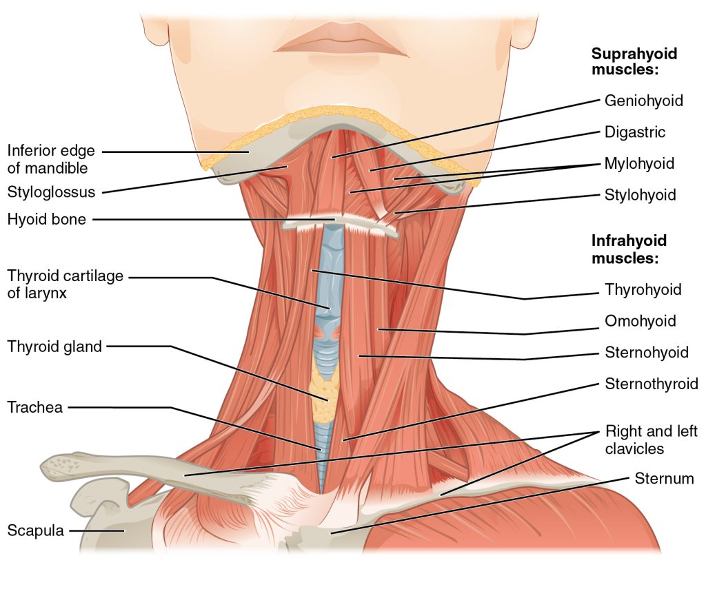
The suprahyoid muscles raise the hyoid bone, the floor of the mouth, and the larynx during deglutition. These include the digastric muscle, which has anterior and posterior bellies that work to elevate the hyoid bone and larynx when one swallows; it also depresses the mandible. The stylohyoid muscle moves the hyoid bone posteriorly, elevating the larynx, and the mylohyoid muscle lifts it and helps press the tongue to the top of the mouth. The geniohyoid depresses the mandible in addition to raising and pulling the hyoid bone anteriorly.
The strap-like infrahyoid muscles generally depress the hyoid bone and control the position of the larynx. The omohyoid muscle, which has superior and inferior bellies, depresses the hyoid bone in conjunction with the sternohyoid and thyrohyoid muscles. The thyrohyoid muscle also elevates the larynx’s thyroid cartilage, whereas the sternothyroid depresses it to create different tones of voice.
Muscles That Move the Head and Back
The head, attached to the top of the vertebral column, is balanced, moved, and rotated by the neck muscles. When these muscles act unilaterally, the head rotates. When they contract bilaterally, the head flexes or extends. The major muscle that laterally flexes and rotates the head is the sternocleidomastoid. In addition, both muscles working together are the flexors of the head. Place your fingers on both sides of the neck and turn your head to the left and to the right. You will feel the movement originate there. This muscle divides the neck into anterior and posterior triangles when viewed from the side (Figure 7.9).
The posterior muscles of the neck are primarily concerned with head movements, like extension. The back muscles stabilize and move the vertebral column, and are grouped according to the lengths and direction of the fascicles.
The splenius muscles originate at the midline and run laterally and superiorly to their insertions. From the sides and the back of the neck, the splenius capitis inserts onto the head region, and the splenius cervicis extends onto the cervical region. These muscles can extend the head, laterally flex it, and rotate it.
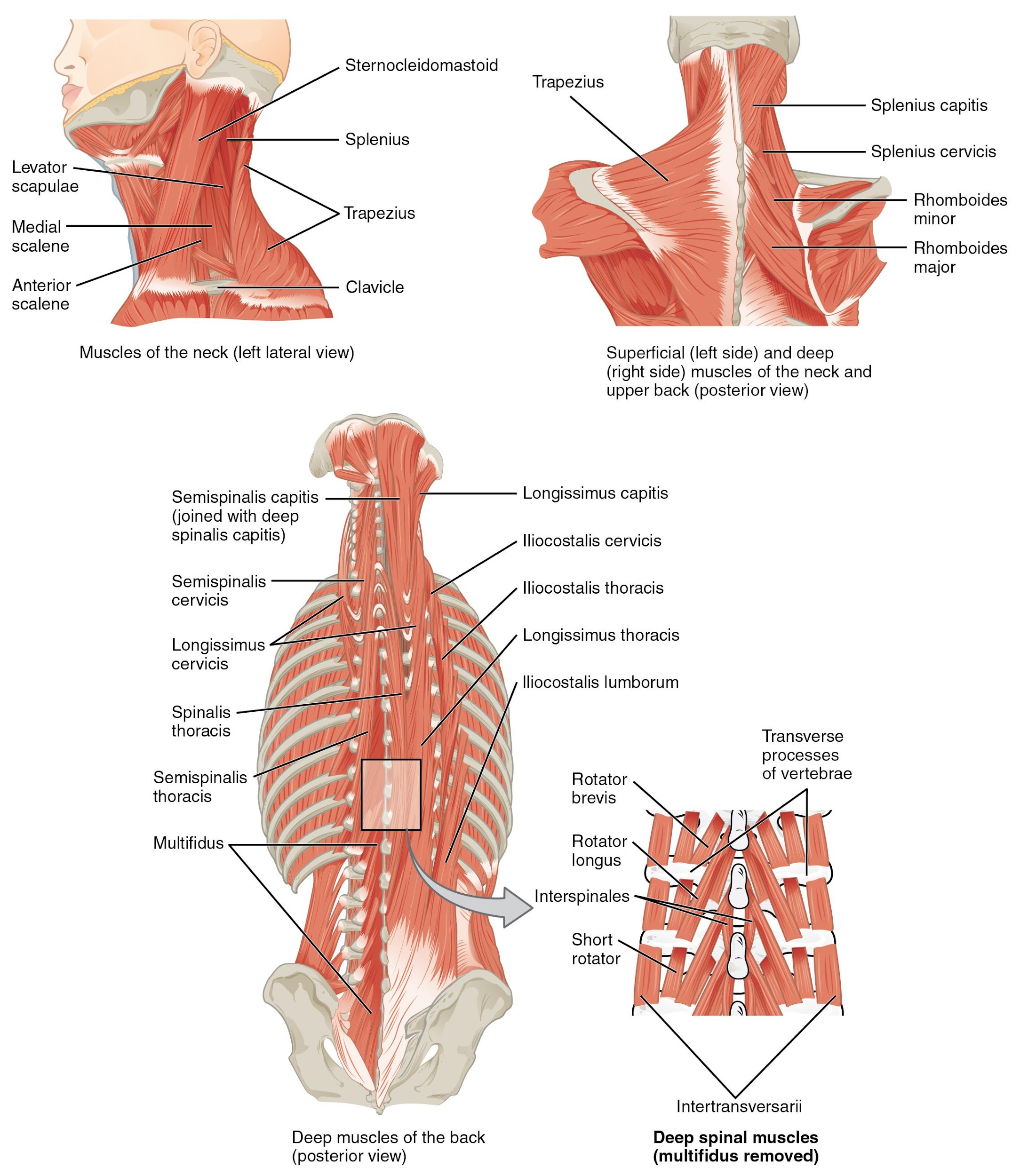
The erector spinae group forms the majority of the muscle mass of the back and it is the primary extensor of the vertebral column. It controls flexion, lateral flexion, and rotation of the vertebral column, and maintains the lumbar curve. The erector spinae comprises the iliocostalis (laterally placed) group, the longissimus (intermediately placed) group, and the spinalis (medially placed) group.
The iliocostalis group includes the iliocostalis cervicis, associated with the cervical region; the iliocostalis thoracis, associated with the thoracic region; and the iliocostalis lumborum, associated with the lumbar region. The three muscles of the longissimus group are the longissimus capitis, associated with the head region; the longissimus cervicis, associated with the cervical region; and the longissimus thoracis, associated with the thoracic region. The third group, the spinalis group, comprises the spinalis capitis (head region), the spinalis cervicis (cervical region), and the spinalis thoracis (thoracic region).
The transversospinales muscles run from the transverse processes to the spinous processes of the vertebrae. Similar to the erector spinae muscles, the semispinalis muscles in this group are named for the areas of the body with which they are associated. The semispinalis muscles include the semispinalis capitis, the semispinalis cervicis, and the semispinalis thoracis. The multifidus muscle of the lumbar region helps extend and laterally flex the vertebral column.
Important in the stabilization of the vertebral column is the segmental muscle group, which includes the interspinales and intertransversarii muscles. These muscles bring together the spinous and transverse processes of each consecutive vertebra. Finally, the scalene muscles work together to flex, laterally flex, and rotate the head. They also contribute to deep inhalation. The scalene muscles include the anterior scalene muscle (anterior to the middle scalene), the middle scalene muscle (the longest, intermediate between the anterior and posterior scalenes), and the posterior scalene muscle (the smallest, posterior to the middle scalene).
Muscles of the Abdomen
It is a complex job to balance the body on two feet and walk upright. The muscles of the vertebral column, thorax, and abdominal wall extend, flex, and stabilize different parts of the body’s trunk. The deep muscles of the core of the body help maintain posture as well as carry out other functions. The brain sends out electrical impulses to these various muscle groups to control posture by alternate contraction and relaxation. This is necessary so that no single muscle group becomes fatigued too quickly. If any one group fails to function, body posture will be compromised.
There are four pairs of abdominal muscles that cover the anterior and lateral abdominal region and meet at the anterior midline. These muscles of the anterolateral abdominal wall can be divided into four groups: the external obliques, the internal obliques, the transversus abdominis, and the rectus abdominis (Figure 7.10).
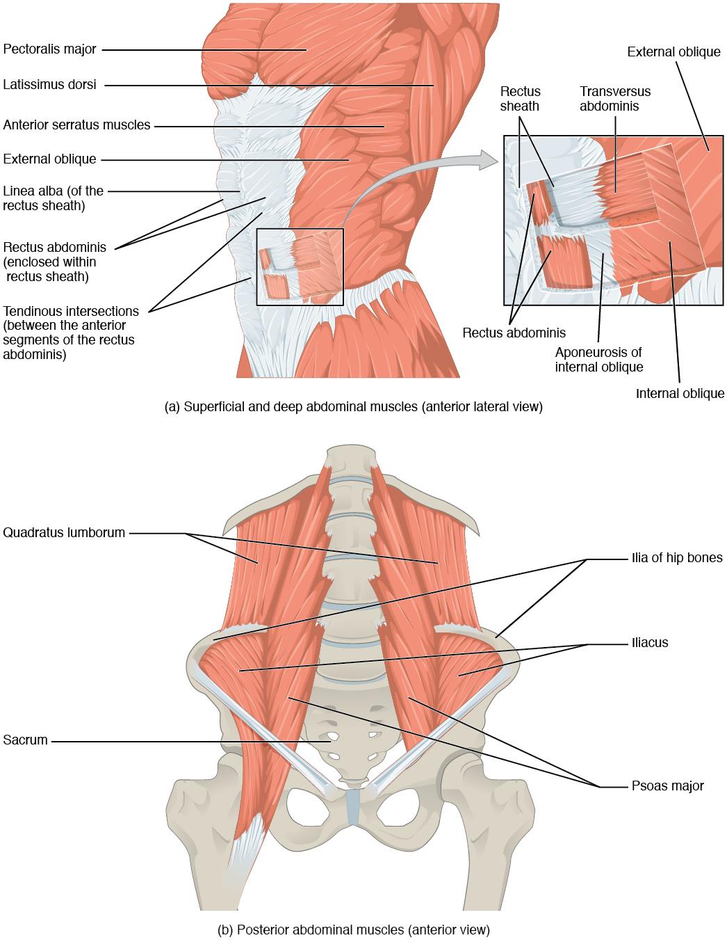
There are three flat skeletal muscles in the antero-lateral wall of the abdomen. The external oblique, closest to the surface, extend inferiorly and medially, in the direction of sliding one’s four fingers into pants pockets. Perpendicular to it is the intermediate internal oblique, extending superiorly and medially, the direction the thumbs usually go when the other fingers are in the pants pocket. The deep muscle, the transversus abdominis, is arranged transversely around the abdomen, similar to the front of a belt on a pair of pants. This arrangement of three bands of muscles in different orientations allows various movements and rotations of the trunk. The three layers of muscle also help to protect the internal abdominal organs in an area where there is no bone.
The linea alba is a white, fibrous band that is made of the bilateral rectus sheaths that join at the anterior midline of the body. These enclose the rectus abdominis muscles (a pair of long, linear muscles, commonly called the “sit-up” muscles) that originate at the pubic crest and symphysis, and extend the length of the body’s trunk. Each muscle is segmented by three transverse bands of collagen fibers called the tendinous intersections. This results in the look of “six-pack abs,” as each segment hypertrophies on individuals at the gym who do many sit-ups.
The posterior abdominal wall is formed by the lumbar vertebrae, parts of the ilia of the hip bones, psoas major and iliacus muscles, and quadratus lumborum muscle. This part of the core plays a key role in stabilizing the rest of the body and maintaining posture.
Muscles of the Thorax
The muscles of the chest serve to facilitate breathing by changing the size of the thoracic cavity. When you inhale, your chest rises because the cavity expands. Alternately, when you exhale, your chest falls because the thoracic cavity decreases in size.
The Diaphragm
The change in volume of the thoracic cavity during breathing is due to the alternate contraction and relaxation of the diaphragm (Figure 7.11). It separates the thoracic and abdominal cavities, and is dome-shaped at rest. The superior surface of the diaphragm is convex, creating the elevated floor of the thoracic cavity. The inferior surface is concave, creating the curved roof of the abdominal cavity.
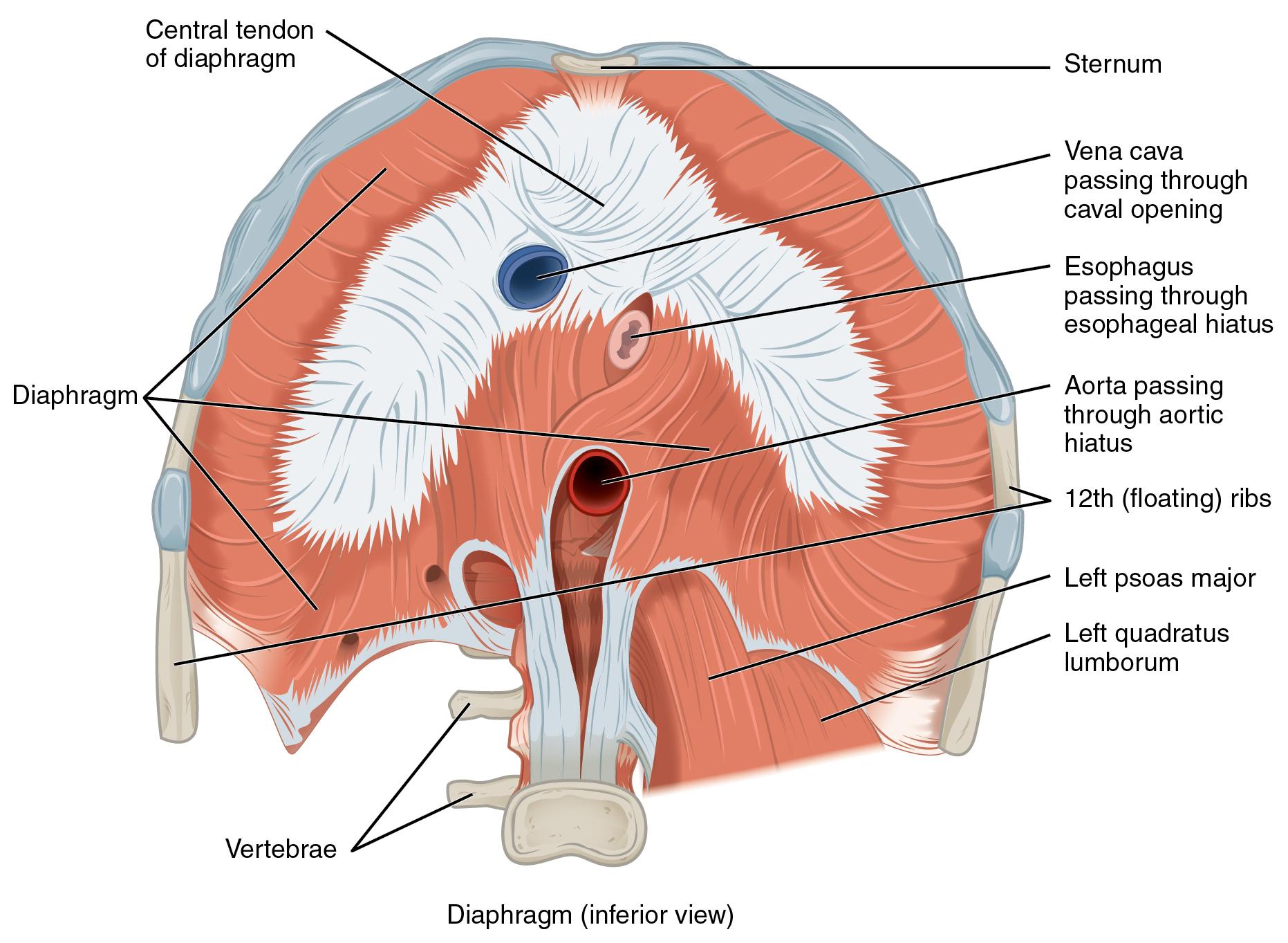
Defecating, urination, and even childbirth involve cooperation between the diaphragm and abdominal muscles (this cooperation is referred to as the “Valsalva maneuver”). You hold your breath by a steady contraction of the diaphragm; this stabilizes the volume and pressure of the peritoneal cavity. When the abdominal muscles contract, the pressure cannot push the diaphragm up, so it increases pressure on the intestinal tract (defecation), urinary tract (urination), or reproductive tract (childbirth).
The inferior surface of the pericardial sac and the inferior surfaces of the pleural membranes (parietal pleura) fuse onto the central tendon of the diaphragm. To the sides of the tendon are the skeletal muscle portions of the diaphragm, which insert into the tendon while having a number of origins including the xiphoid process of the sternum anteriorly, the inferior six ribs and their cartilages laterally, and the lumbar vertebrae and 12th ribs posteriorly.
The diaphragm also includes three openings for the passage of structures between the thorax and the abdomen. The inferior vena cava passes through the caval opening, and the esophagus and attached nerves pass through the esophageal hiatus. The aorta, thoracic duct, and azygous vein pass through the aortic hiatus of the posterior diaphragm.
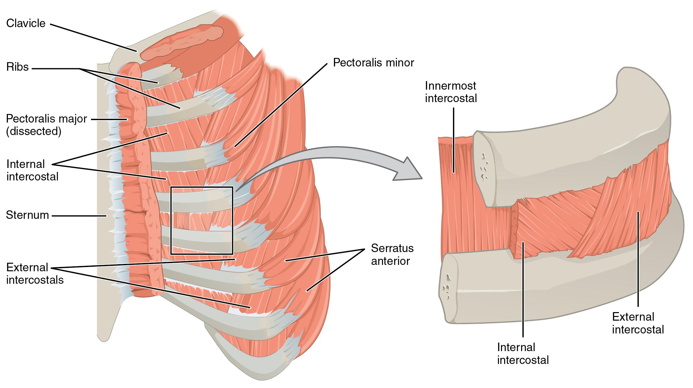
The Intercostal Muscles
There are three sets of muscles, called intercostal muscles, which span each of the intercostal spaces. The principal role of the intercostal muscles is to assist in breathing by changing the dimensions of the rib cage (Figure 7.12).
The 11 pairs of superficial external intercostal muscles aid in inspiration of air during breathing because when they contract, they raise the rib cage, which expands it. The 11 pairs of internal intercostal muscles, just under the externals, are used for expiration because they draw the ribs together to constrict the rib cage. The innermost intercostal muscles are the deepest, and they act as synergists for the action of the internal intercostals.
Unless otherwise indicated, this chapter contains material adapted from Anatomy and Physiology 2e (on OpenStax), by Betts, et al. and is used under a a CC BY 4.0 international license. Download and access OpenStax Anatomy and Physiology for free at https://openstax.org/details/books/anatomy-and-physiology-2e/
