
9 Vascular Tissues & Plant Functional Anatomy
Following this lab, students will be able to:
- Identify the different cell types of plant vascular tissue and describe how they form the overall tissue structure
- Explain how water and sugar are transported in plants
- Identify the main types of cells and tissues found in plants and describe their distribution in roots, stems, and leaves
- Compare and contrast the arrangement of tissues between different parts of a plant (roots, stems, and leaves) and different phylogenetic groupings of plants
- Describe how the cells and tissues of a plant work together to perform the plant’s necessary functions
Contribution Points:
 Consult with your TA to receive a stamp at the end of your lab period.
I have completed the necessary tasks required during this week’s lab to earn Contribution Points. I am aware that I may have point(s) deducted from my Contribution Points if my workspace is not appropriately clean at the conclusion of lab.
Resources
- Compound Scope Tutorial (video tutorial, linked on Canvas)
- Stereomicroscope Tutorial (video tutorial, linked on Canvas)
- How to use Preview (video tutorial, linked on Canvas)
- Using Leica Software (video tutorial, linked on Canvas)
- Biological Science (8th ed.), Freeman et al., 2024. Chapter 34.3–34.4 (pp. 754–763) and Chapter 35 (pp. 766–784).
- Botany Photo Atlas (provided for use in lab)
- Canvas Resources
Background
This week’s lab is a continuation from last week’s investigation of Plant Cells and Tissues. The focus of this week’s lab will be two-fold. First, you will continue studying Plant Cells and Tissues by investigating Plant Vascular Tissues and water transport in plants. You will then take a broader view of plants by considering the plant as a whole and how all of the cells and tissues work together to keep the plant alive.
You’ll be using your knowledge of plant cells and tissues to think about how they are utilized within the plant; how the cells and tissue types work together to form the plant and to perform the plant’s necessary functions.
In lab you will be looking at plants from the perspective of the three basic plant organs; roots, stems, and leaves. Each of these plant organs performs specific tasks for the plant body as a whole, so each organ will display anatomy that allows the plant to fulfill its function. As you go through this week’s material, think about what function each plant organ is responsible for and think about how the anatomy you observe helps to serve that function.
Plant Vascular Tissue
The vascular system enables the transport of fluids and solutes through the plant, similar to the basic function of the cardiovascular system in animals. The vascular system in plants can also provide some support to the plant. It has two main types of tissue: xylem and phloem. Xylem conducts water and dissolved nutrients from roots to shoots through long columns of tissue. Within the tissue there are specialized cells known as tracheids and vessel elements that participate in transport, and parenchyma cells that provide structural support. Tracheids and vessel elements die at maturity and are then able to conduct water through the plant as long columns of hollowed-out dead cells.
Tracheids are long, slender cells with tapered ends. They have gaps in their secondary cell wall that appear as pits. The water moves between tracheids through these pits. Besides water conduction, tracheids can also provide support to the plant. Vessel elements are shorter and wider than tracheids. Along with pits, they also have perforations in their end walls, allowing the vessel elements to be stacked up along each other like a tube. The perforations in vessels are gaps in both the primary and secondary cell walls.
Phloem tissue is made up of two types of specialized cells: sieve-tube elements and companion cells. Sieve tube elements are long, thin cells that can stack together in a tube, similar to xylem cells. They have perforated ends called sieve plates, which allow sugars and other organic materials to pass between the cells and throughout the plant body. The sieve tube elements are considered living at maturity, but they lack nuclei, chloroplasts and many other organelles. They are connected to companion cells via plasmodesmata. Companion cells contain most organelles and support the sieve tube elements, performing many of their necessary metabolic processes. Phloem tissue also contains more generalized parenchyma cells that provide structural support.
The majority of the specimens you will be observing in lab are provided to you as prepared slides. Not to prepare your own slides (for most specimens) will save you some time this week.
You will be utilizing the microscopes and digital cameras in lab this week. Be mindful when labeling and managing your images to ensure that you do not lose your images or mislabel your photos.
TA Demonstration
Investigating Water Movement in Bok Choy Petioles
Your TA will play a video tutorial demonstration of water movement in bok choy petioles. In this demonstration, two bok choy leaves (one with the leaves intact, and one where the leaf blades were removed) are cut through the bases of the petioles and placed immediately into a dye solution. The petioles stay in the dye for a short time before they are removed and the movement of the dye through the petioles is examined.
![]() How does the presence of leaf blades affect the rate of water movement through the xylem?
How does the presence of leaf blades affect the rate of water movement through the xylem?
![]() Is this consistent with the evaporation-cohesion-tension model?
Is this consistent with the evaporation-cohesion-tension model?
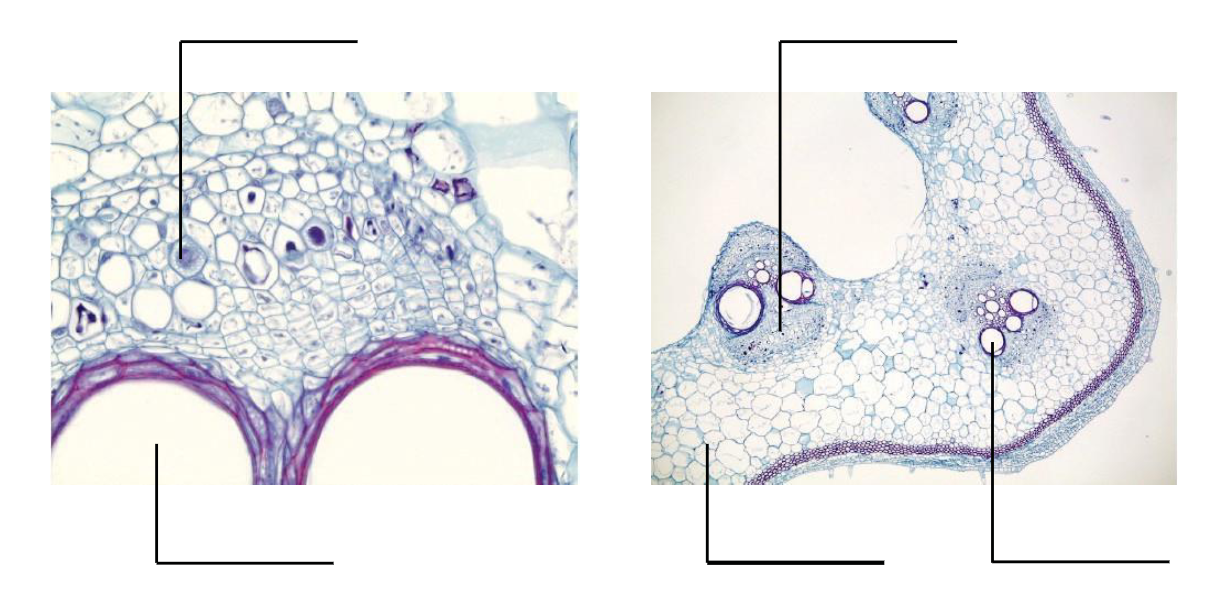
1. Xylem – Tracheids
Specimen: Prepared slide of pine stem secondary xylem.
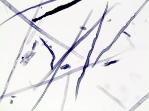
![]() What kind(s) of vascular cells can you identify on this slide?
What kind(s) of vascular cells can you identify on this slide?
![]() What are the identifying characteristics of these cells?
What are the identifying characteristics of these cells?
2. Xylem – Tracheids and Vessel Elements
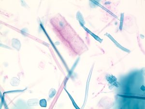
Specimen: Prepared slide of pumpkin stem macerate.
![]() What kind(s) of vascular cells can you identify on this slide?
What kind(s) of vascular cells can you identify on this slide?
COMPARISON OF SPECIMEN 1 AND SPECIMEN 2
Draw a tracheid in the circle on the left.
Draw a vessel element in the circle on the right.
![]() Which specimen contains both xylem cell types?
Which specimen contains both xylem cell types?
![]() What are the structural and/or functional differences between vessel elements and tracheids?
What are the structural and/or functional differences between vessel elements and tracheids?
![]() Do these cells contain cytoplasm and/or a nucleus at functional maturity?
Do these cells contain cytoplasm and/or a nucleus at functional maturity?
![]() What are the functions of vessel elements and tracheids?
What are the functions of vessel elements and tracheids?
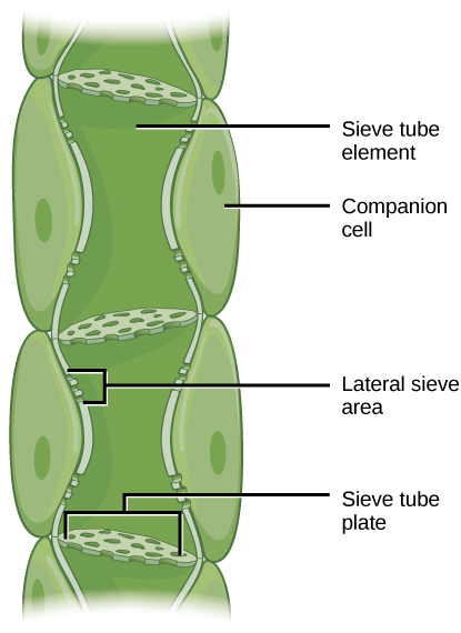
3. Phloem Tissues – Sieve Tube Elements and Companion Cells
Specimen: Prepared slide of pumpkin stem longitudinal section. Label parenchyma, sieve tube member, companion cell, and sieve tube plate on the image below.
![]() What cell types would you find in the phloem tissue?
What cell types would you find in the phloem tissue?
![]() What are the functions of these cells?
What are the functions of these cells?
![]() Do phloem cells contain cytoplasm and/or a nucleus at functional maturity?
Do phloem cells contain cytoplasm and/or a nucleus at functional maturity?
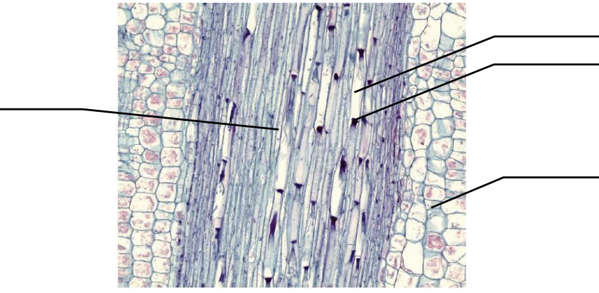
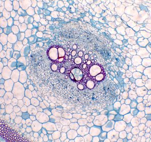
4. Xylem and Phloem in Prepared Cross Section
Specimen: Prepared slide of pumpkin stem cross section.
![]() Locate and label the xylem and phloem tissues in images of this cross section below.
Locate and label the xylem and phloem tissues in images of this cross section below.
![]() Locate and label a sieve plate in images of this cross section below.
Locate and label a sieve plate in images of this cross section below.
COMPARISON OF SPECIMEN 3 AND SPECIMEN 4
These two specimens give you different views of the same plant. Compare what you see in these two specimens to give you a better idea of how the vascular tissue cells are shaped.
![]() Describe the shapes and functions of the following cell types:
Describe the shapes and functions of the following cell types:
Vessel Element
Sieve Tube Elements
Companion Cell
Xylem and Phloem in Fresh Cross Section
Specimen: Prepare a wet mount slide of a sunflower (Helianthus) or tomato (Solanum) in both cross section and longitudinal section.
Locate the xylem and phloem in your cross section.
![]() What is the function of xylem?
What is the function of xylem?
![]() What is the function of phloem?
What is the function of phloem?
ROOTS
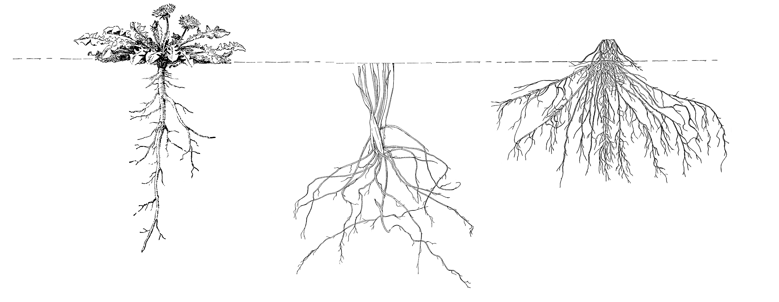
6. Root Morphology
The figure below illustrates the three major types of roots found in plants; tap roots, fibrous roots, and adventitious roots.
![]() In your own words, describe the differences between the three types of roots below.
In your own words, describe the differences between the three types of roots below.
7. Radish Seedling Observations ( shoot, root, root hairs, root cap)
Specimen: Make observations of the radish seedlings from the plate provided.
![]() Locate the shoot, root, and root hairs on the radish seedlings. What is the function of the root hairs?
Locate the shoot, root, and root hairs on the radish seedlings. What is the function of the root hairs?
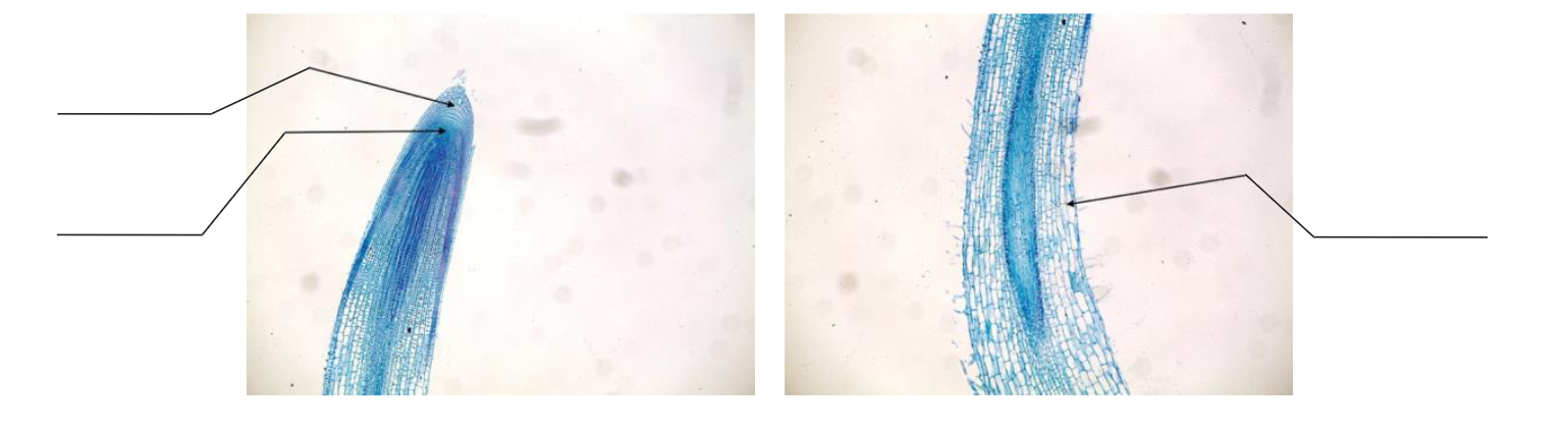
8. Root Anatomy (root cap, region of cell division, region of cell elongation)
Specimen: Prepared slide of longitudinal section of young dicot root (radish, Raphanus).
![]() Locate the root cap, region of cell division, and region of elongation in the images below.
Locate the root cap, region of cell division, and region of elongation in the images below.
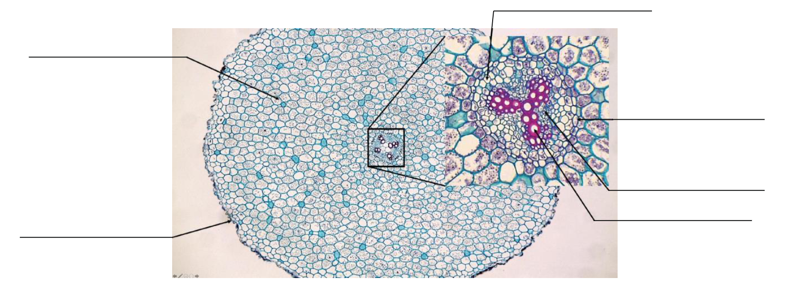
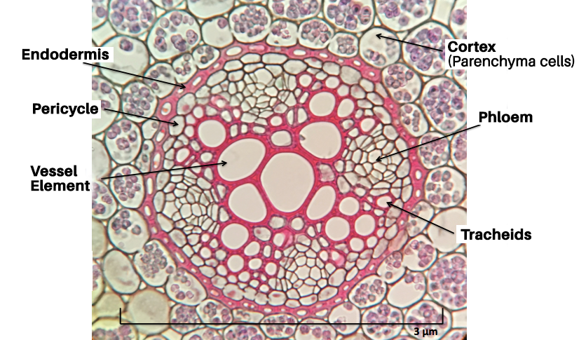
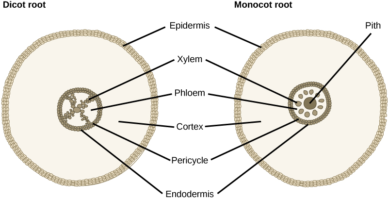
9. Eudicot Root Anatomy
Specimen: Eudicot Roots—Prepared slide of buttercup (Ranunculus) cross section. Examine the slide carefully under the compound microscope.
![]() Label the following structures on the image below: epidermis, cortex, xylem, phloem, pericycle, and endodermis.
Label the following structures on the image below: epidermis, cortex, xylem, phloem, pericycle, and endodermis.
![]() What is the purpose of the Casparian Strip?
What is the purpose of the Casparian Strip?
![]() Can you see the vascular cambium, secondary xylem, secondary phloem, and cork cambium in this slide? Why or why not?
Can you see the vascular cambium, secondary xylem, secondary phloem, and cork cambium in this slide? Why or why not?
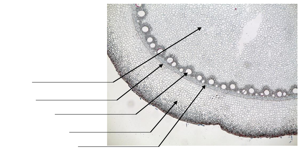
10. Monocot Root Anatomy
Specimen: Monocot Roots— Prepared slide of cross section of corn (Zea mays) root (typical monocot root).
![]() Examine the organization of the vascular tissue of the corn root. How does it differ from a eudicot root?
Examine the organization of the vascular tissue of the corn root. How does it differ from a eudicot root?
![]() Label the following structures on the photo below: pith, endodermis, cortex, xylem, and phloem.
Label the following structures on the photo below: pith, endodermis, cortex, xylem, and phloem.
Stems
11. External Eudicot Stem Structure
Specimen: Woody twig displays (various local tree species)
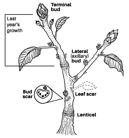
Locate the apical buds, lateral buds, leaf scars, and lenticels on the specimens provided in lab.
![]() Why is bark important to woody stems?
Why is bark important to woody stems?
![]() What function do the lenticels perform?
What function do the lenticels perform?
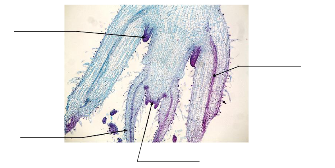
12. Herbaceous (Non- Woody) Eudicot Stem Histology
Specimen: Prepared slide of longitudinal section of Coleus terminal bud.
![]() Locate the leaf primordium, vascular bundle, apical meristem, and lateral bud on the photo below.
Locate the leaf primordium, vascular bundle, apical meristem, and lateral bud on the photo below.
![]() What is the difference between the terminal bud and the lateral bud? What is similar?
What is the difference between the terminal bud and the lateral bud? What is similar?
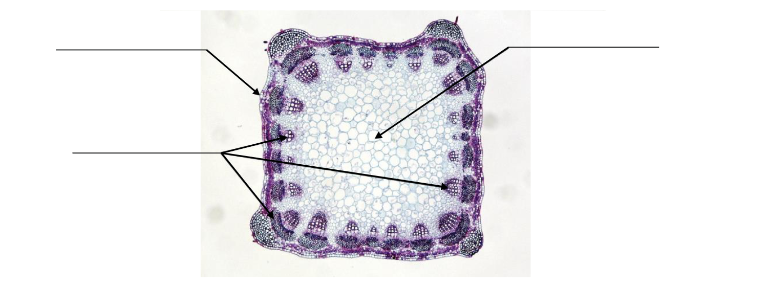
13. Eudicot Stem Fresh Cross Section
Specimen: Prepare your own cross section of sunflower (Helianthus) or tomato (Solanum) stem.
![]() Carefully examine the slide and locate the pith, epidermis, and vascular bundles on the photo below.
Carefully examine the slide and locate the pith, epidermis, and vascular bundles on the photo below.
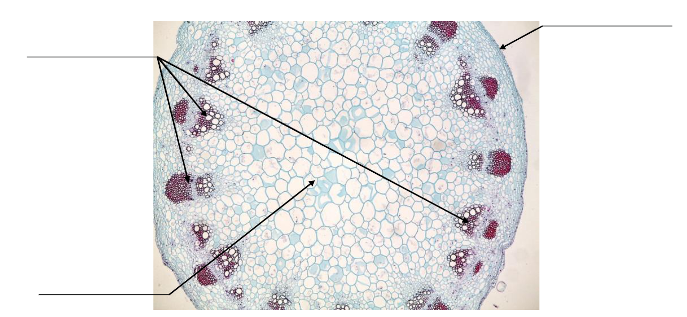
14. Eudicot Stem Cross Section Prepared Slide
Specimen: Prepared slide of alfalfa (Medicago) stem cross section.
![]() Carefully examine the prepared slide and locate the pith, epidermis, and vascular bundles on the image below.
Carefully examine the prepared slide and locate the pith, epidermis, and vascular bundles on the image below.
15. Monocot Stem Histology
Specimen: Prepared slide of corn (Zea mays) stem cross section.
![]() Carefully examine the prepared slide and locate the ground tissue (parenchyma), epidermis, and vascular bundles on the image on the next page.
Carefully examine the prepared slide and locate the ground tissue (parenchyma), epidermis, and vascular bundles on the image on the next page.
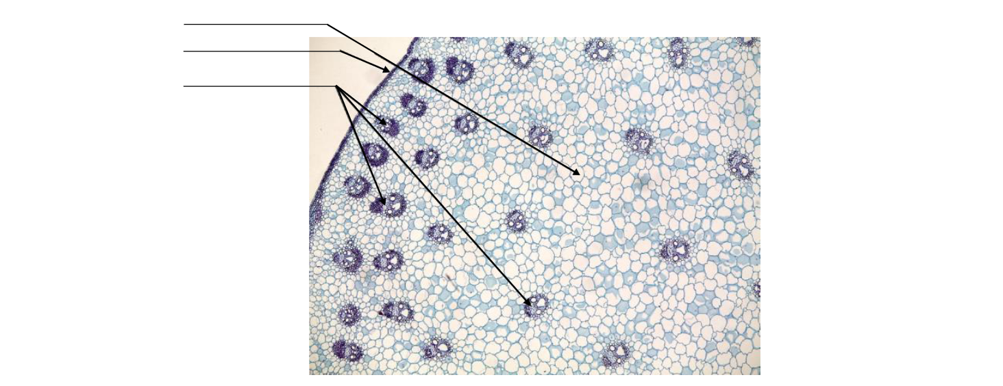
![]() Referring to Figure 9.18, how is the organization of the vascular tissue in monocot stems different from that in eudicot stems?
Referring to Figure 9.18, how is the organization of the vascular tissue in monocot stems different from that in eudicot stems?
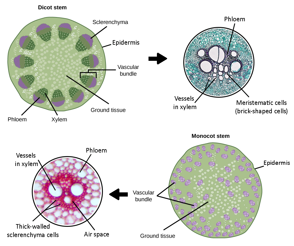
Review of vascular tissue placement in monocot and eudicot stems and roots. Draw a line to match the options on the right to the statements on the left to complete them:
Vascular tissue in monocot roots is:
Vascular tissue in monocot stems is:
Vascular tissue in eudicot roots is:
Vascular tissue in eudicot stems is:
found in the center.
arranged in a ring.
arranged in a ring.
scattered throughout.
Leaves
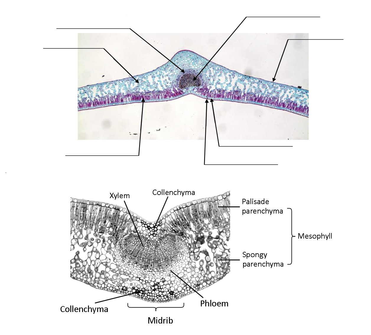
16. Internal Eudicot Leaf Anatomy
Specimen: Structure of eudicot leaf. Prepared slide of cross section of eudicot leaf AND fresh cross section of eudicot leaf.
![]() Review from Laboratory 8: What is a stoma/stomate? What is the function of the guard cells?
Review from Laboratory 8: What is a stoma/stomate? What is the function of the guard cells?
![]() Carefully look at both your own cross section of fresh material and the prepared slide of the eudicot leaf cross section. Describe what you see. Do you notice any differences between the prepared slide and the fresh specimen?
Carefully look at both your own cross section of fresh material and the prepared slide of the eudicot leaf cross section. Describe what you see. Do you notice any differences between the prepared slide and the fresh specimen?
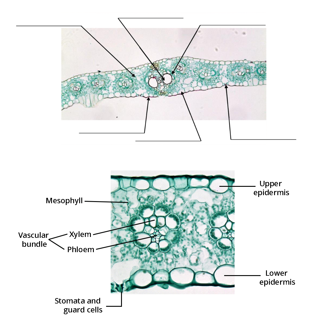
![]() Locate and label the following structures on the photo below: cuticle, epidermis, palisade mesophyll, spongy mesophyll, bundle sheath, vascular bundle, and stoma.
Locate and label the following structures on the photo below: cuticle, epidermis, palisade mesophyll, spongy mesophyll, bundle sheath, vascular bundle, and stoma.
17. Internal Monocot Leaf Anatomy
Specimen: Structure of monocot leaf. Prepared slide of corn (Zea mays) leaf cross section AND fresh cross section of corn leaf (Zea mays).
![]() Carefully look at both your own cross section of fresh material and the prepared slide of the monocot leaf cross section.
Carefully look at both your own cross section of fresh material and the prepared slide of the monocot leaf cross section.
![]() Describe what you see. Do you notice any differences between the prepared slide and the fresh specimen?
Describe what you see. Do you notice any differences between the prepared slide and the fresh specimen?
![]() Locate and label the following structures on the image below: cuticle, epidermis, bundle sheath, vascular tissue, mesophyll, and stoma.
Locate and label the following structures on the image below: cuticle, epidermis, bundle sheath, vascular tissue, mesophyll, and stoma.
18. Internal Anatomy of Gymnosperm Leaf
Specimen: Structure of gymnosperm needle. Prepared slide of cross section of Austrian pine (Pinus nigra) needle.
![]() Carefully examine the slide and describe what you see. What is the general tissue and cell organization of a gymnosperm needle?
Carefully examine the slide and describe what you see. What is the general tissue and cell organization of a gymnosperm needle?
![]() What structure(s) do you see in the gymnosperm leaf cross section that you did not see in the monocot or eudicot leaf cross sections?
What structure(s) do you see in the gymnosperm leaf cross section that you did not see in the monocot or eudicot leaf cross sections?
![]() Do you notice anything special about the stomata in the pine leaf compared to other leaves you observed in lab?
Do you notice anything special about the stomata in the pine leaf compared to other leaves you observed in lab?
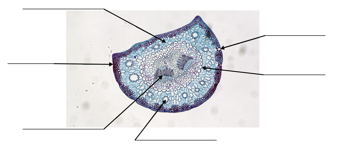
![]() Locate and label the following structures on the image below: epidermis, mesophyll, vascular bundle, stoma, resin duct, bundle sheath.
Locate and label the following structures on the image below: epidermis, mesophyll, vascular bundle, stoma, resin duct, bundle sheath.
![]() Compare the pine needle (gymnosperm leaf) to the leaves of monocots and eudicots. Specifically look at the epidermal thickness, cross-sectional shape, position of vascular tissue, and presence of resin ducts. Describe what you see in the gymnosperm leaf vs. the monocot/eudicot leaves for each of these characteristics.
Compare the pine needle (gymnosperm leaf) to the leaves of monocots and eudicots. Specifically look at the epidermal thickness, cross-sectional shape, position of vascular tissue, and presence of resin ducts. Describe what you see in the gymnosperm leaf vs. the monocot/eudicot leaves for each of these characteristics.
