
10 Digestive, Renal, and Reproductive Systems
Following this lab, students will be able to:
- Demonstrate proper dissection technique and respect for animal specimens and dissection tools
- Identify the structures and describe the functions of the mammalian digestive, renal, and reproductive systems
- Compare and contrast fetal pig anatomy and physiology with that of humans and other mammals
- Predict the consequences of a defect in the anatomy and/or physiology of mammalian systems
- Name the substances that are created by and/or pass through the digestive, renal, and reproductive organ systems and describe the pathway they take through these systems
Contribution Points:
 Consult with your TA to receive a stamp at the end of your lab period.
I have completed the necessary tasks required during this week’s lab to earn Contribution Points. I am aware that I may have point(s) deducted from my Contribution Points if my workspace is not appropriately clean at the conclusion of lab.
Your TA will check that you have attempted to complete the dissections associated with this week’s lab activity.
Resources
- Dissection Interchapter 6 “Dissection Techniques and Terminology” (pp.137–140)
- Fetal pig dissection tutorial video (tutorial video on Canvas)
- Fetal pig dissection guide, Smith and Schenk 2011 (provided for use in lab)
- Biological Science, Freeman et al. 2024 (8th ed.) Digestive System (41.3), Renal System (40.4), Reproductive System (47.2–47.3)
- Internet and Canvas resources
![]() This icon represents a question meant to test your understanding. Answering these questions in the space provided as you go through the lab will help you better understand the topic and study more effectively. Use your text or e-book, pig dissection guide, and the internet to help you.
This icon represents a question meant to test your understanding. Answering these questions in the space provided as you go through the lab will help you better understand the topic and study more effectively. Use your text or e-book, pig dissection guide, and the internet to help you.
1. All About Your Pig
This is your first day of fetal pig dissection. Working with a partner, obtain a pig and prepare your specimen and workstation as shown in the “Fetal pig dissection video tutorial”. Before you begin your dissection, do a thorough investigation of its external features.
![]() Based on your observations of your fetal pig’s external anatomy (specifically the location of its urogenital opening), is your pig a male or female? How can you tell?
Based on your observations of your fetal pig’s external anatomy (specifically the location of its urogenital opening), is your pig a male or female? How can you tell?
After you have determined the sex of your pig, you can begin to make the incisions shown in the next section to expose its internal organs.
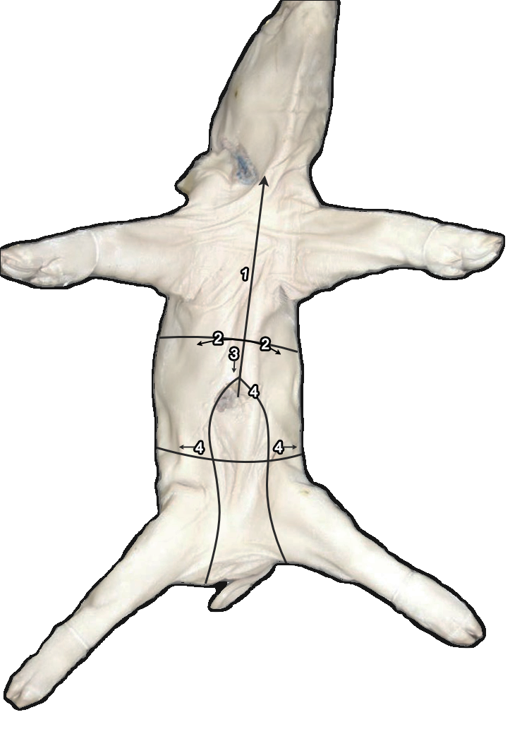
2. Making the First Cuts
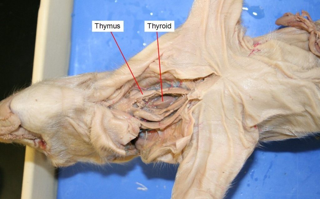
Use Figure 10.1 below to make your first incisions. Follow the numbers to make the incisions in order. Make sure you don’t slice too deep and puncture the organs. It is always better to cut slowly and shallowly to start, then cut deeper later if you need to. After you make the cuts, identify the organs shown in Figure 10.2. BE CAREFUL not to remove the thymus or thyroid gland when you take off the skin on the neck. Leave the esophagus in place.
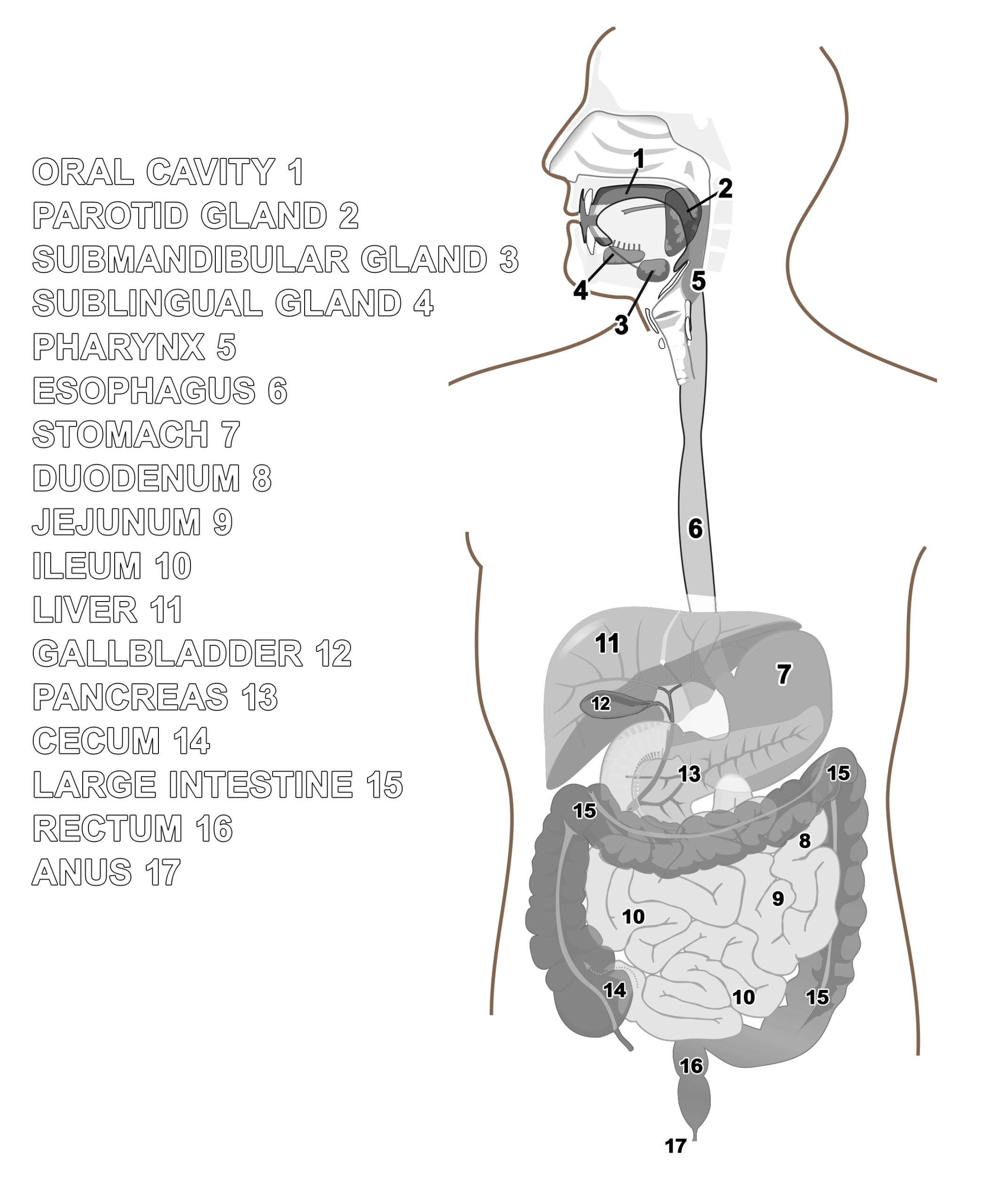
3. Body Cavity and Digestive Tract Anatomy
Locate each organ in your pig shown in Figures 10.2 and 10.3. Have your partner test you for recognition.
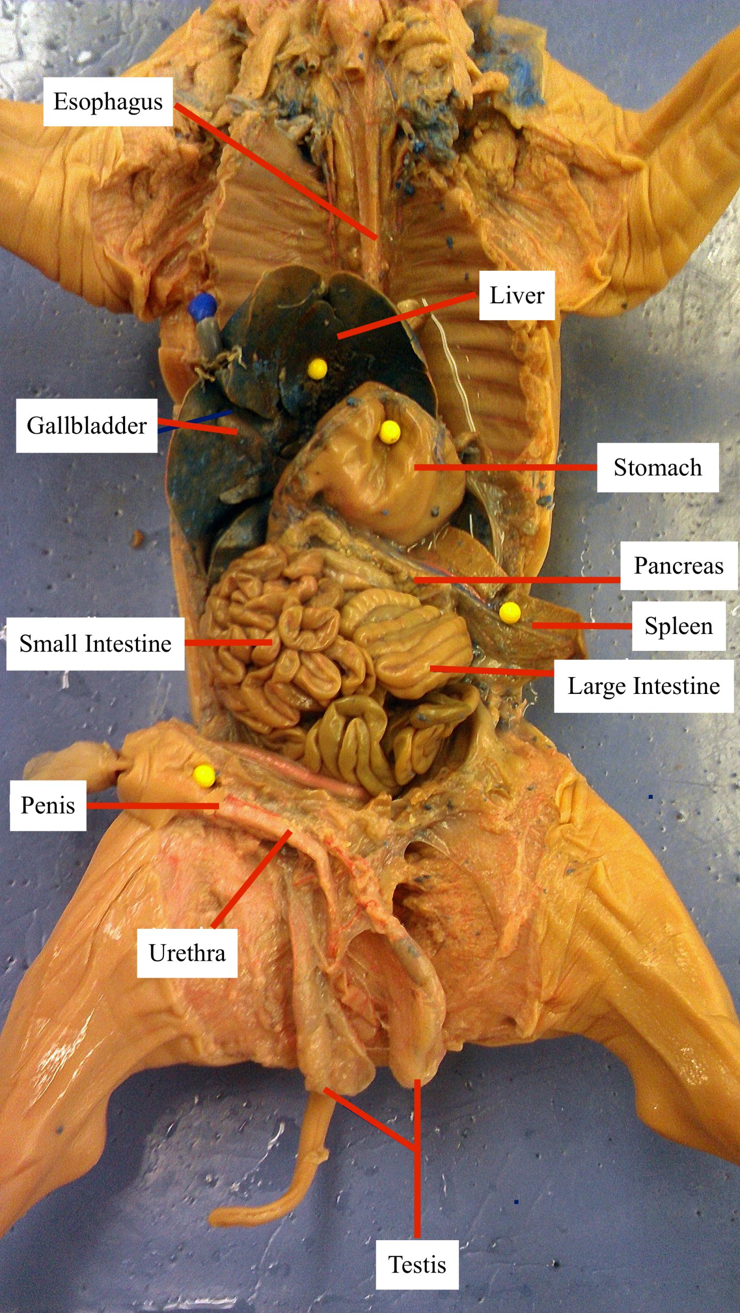
![]() What are the largest organs visible in your fetal pig? What are the functions of these organs and why do you think they are so large?
What are the largest organs visible in your fetal pig? What are the functions of these organs and why do you think they are so large?
One group from each class should remove the intestines from their fetal pig for measuring. Estimate or measure the length of the removed small and large intestines.
Intestine Length =
![]() Did the length of your fetal pig’s intestines surprise you? Why do you think the intestines are so long?
Did the length of your fetal pig’s intestines surprise you? Why do you think the intestines are so long?
 After you have thoroughly investigated your fetal pig’s intestines, go to the Prepared Slide of Jejunum (intestines) Station and observe the prepared microscopic slides of the small intestine. Make sure you locate the villus, microvilli, lumen, and smooth muscle. Sketch what you see in the adjacent circle and/or take a photo using the Leica software.
![]() How do the villi and microvilli relate to the intestine’s function?
How do the villi and microvilli relate to the intestine’s function?
![]() Celiac disease occurs in some individuals when ingested gluten causes an autoimmune response. This response often causes damage to the small intestine, including shortening of the villi. Symptoms of celiac disease include diarrhea, weight loss, anemia, and other mineral and nutrient deficiencies. Why would damage to the villi cause these symptoms?
Celiac disease occurs in some individuals when ingested gluten causes an autoimmune response. This response often causes damage to the small intestine, including shortening of the villi. Symptoms of celiac disease include diarrhea, weight loss, anemia, and other mineral and nutrient deficiencies. Why would damage to the villi cause these symptoms?
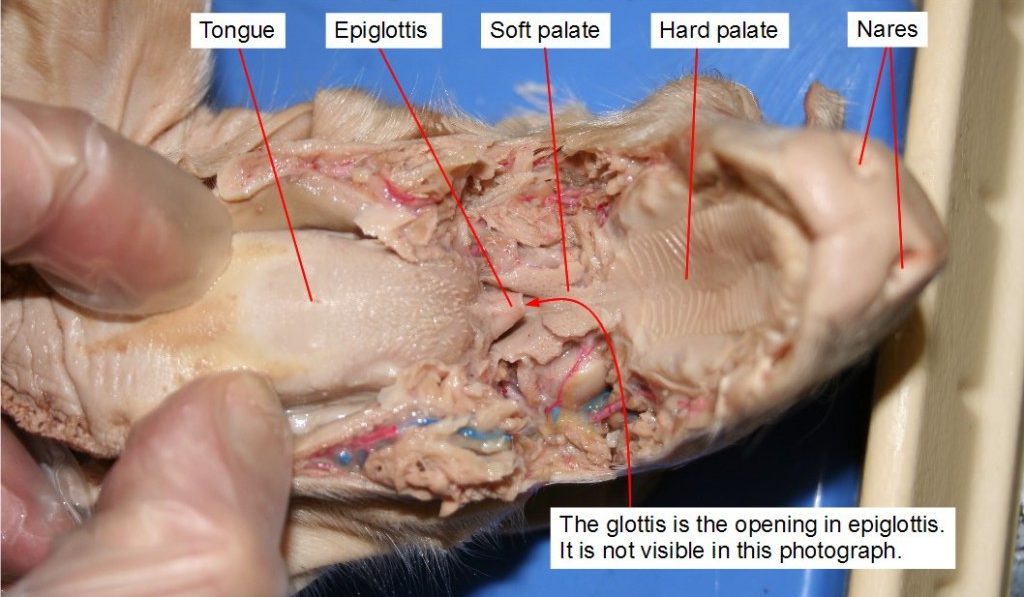
4. Esophagus, Trachea, and Mouth Anatomy
When you are working to uncover the esophagus, trachea, and larynx, BE CAREFUL not to cut any blood vessels. We will need these intact for next week’s lab. Use Figure 10.2 and Figure 10.3 to help you locate the esophagus in your pig. For help in locating the trachea and larynx, look ahead to Figure 12.2 in the Gas Exchange chapter for a detailed image.
![]() What are the functions of these structures? Fill in the table below.
What are the functions of these structures? Fill in the table below.
| Structure | Function |
| Esophagus | |
| Trachea | |
| Salivary Glands |
The mouth anatomy of mammals is quite complex and dissection of this area can be complicated. Demonstration dissections have been provided for you, but you may choose to do your own dissection of the mouth and salivary glands if you would like a challenge and have time. Using the demo dissections or your own, locate the mouth anatomy structures shown in Figure 10.4 of the fetal pig.
5. Accessory Organs
Be sure to locate the liver, gallbladder, pancreas, spleen, thyroid, and thymus. Refer to Figures 10.2, 10.3, and 10.5 to help you. Note that the thyroid is an endocrine gland and the thymus is part of the lymphatic system, but because we don’t study those systems separately, we will examine them here. What are their functions?
| Structure | Function |
| Salivary Glands | |
| Liver | |
| Gallbladder | |
| Pancreas | |
| Spleen | |
| Thyroid | |
| Thymus |
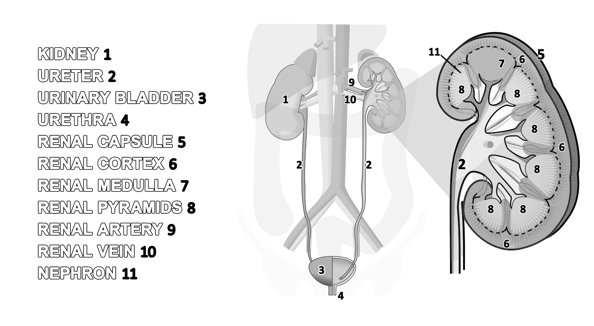
6. Mammalian Excretory System
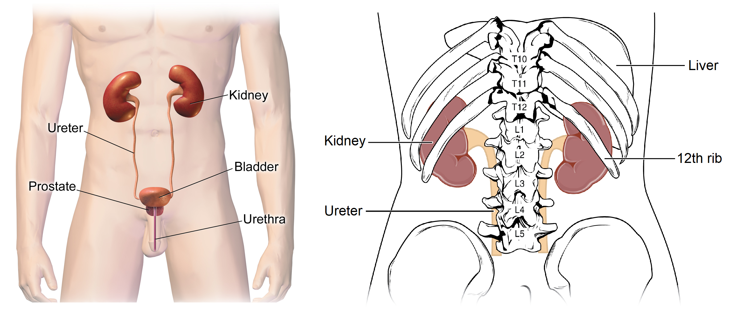
The excretory system is extremely important in mammals to help eliminate waste, filter materials, and regulate water and salt levels. Blood is continually filtered through the kidney; every time the heart beats, about 20% of the blood is routed through the kidneys. Plasma and solutes leak out of capillaries in the kidneys and is then modified into urine. Once created, urine travels through the ureters, the bladder, the urethra, and then exits the body. Identify the components of the excretory system as shown in Figure 10.6 in your dissected fetal pig and then we will explore these in more detail.
After you have located all the structures above, remove one kidney from your fetal pig. Take it to the Kidney Station and cut a thin section from it using a razor blade. Find the cortical and medullary regions. Compare your section to the prepared kidney cross section slide available to you at the station.
The kidney is very complex; use the kidney model and poster in the lab to explore the internal structure of the kidneys that you cannot see with your naked eye. Utilizing the model while viewing the prepared slides of the kidney cross section may help you interpret what you see under the scope.
![]() Based on your observation of the kidney model, what do you think is the functional unit of the kidney? Why?
Based on your observation of the kidney model, what do you think is the functional unit of the kidney? Why?
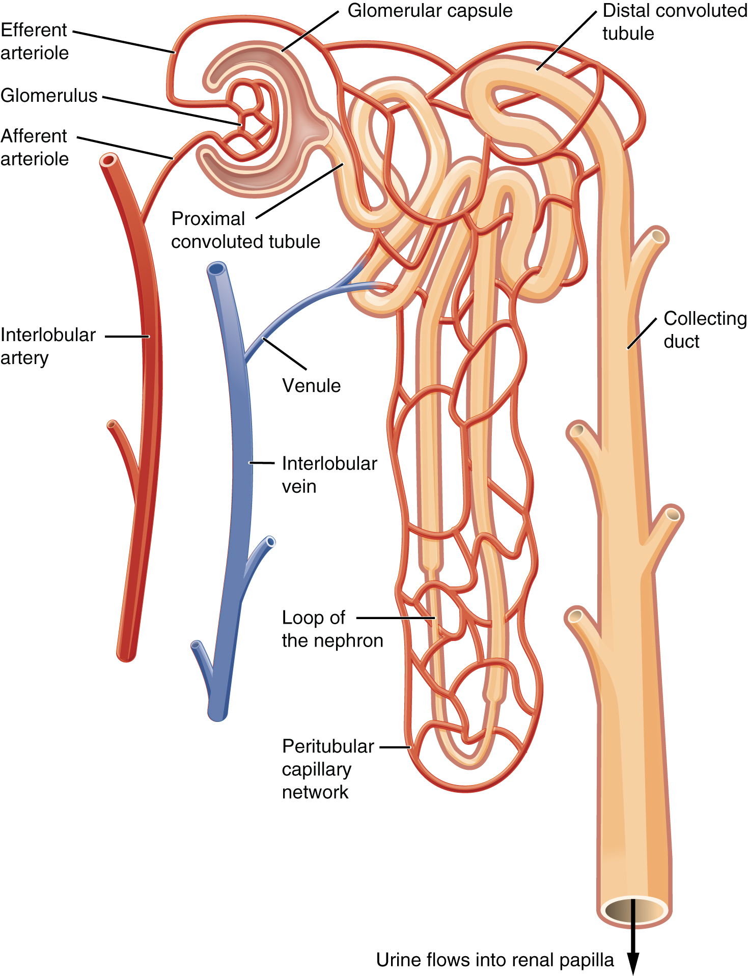
Locate the following kidney and nephron structures in Figure 10.6 using the model and provided slides.
![]() Why do you think the arteries leading to the kidneys are so large?
Why do you think the arteries leading to the kidneys are so large?
![]() What do the kidneys filter from the blood?
What do the kidneys filter from the blood?
![]() Trace the flow of fluid through a nephron from the glomerulus to the collecting duct in Figure 10.7 using your pencil. What is the order of structures through which the fluid passes?
Trace the flow of fluid through a nephron from the glomerulus to the collecting duct in Figure 10.7 using your pencil. What is the order of structures through which the fluid passes?
7. Mammalian Reproductive System
Identify the internal reproductive organs of your pig. This may take some careful dissection to prevent accidentally cutting through structures. Locate a group of your classmates or carefully observe the demonstration dissection of a pig of the opposite sex and make sure you can identify both male and female reproductive organs, as shown in Figures 10.8 and 10.9.
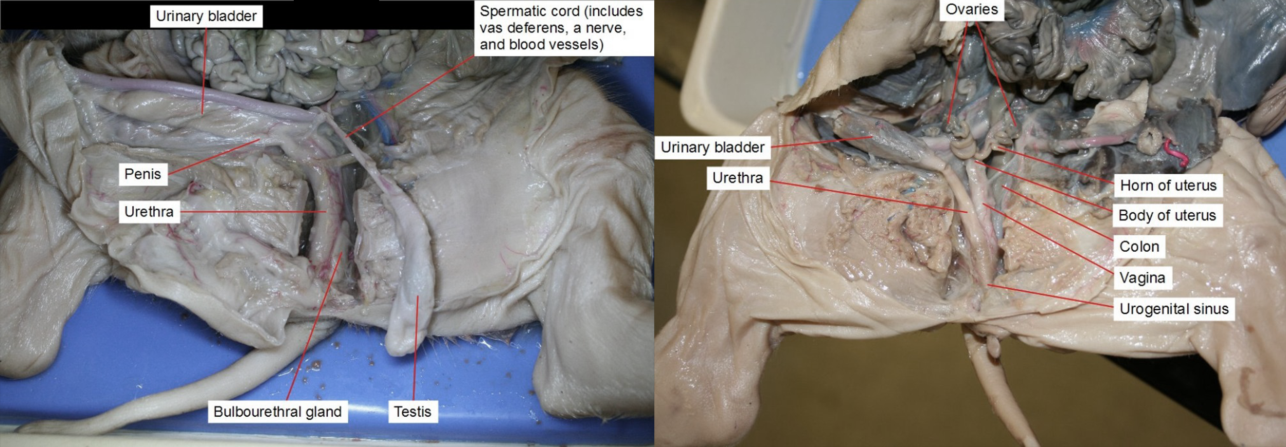
Describe the pathway that eggs and sperm take to leave the body.
Eggs:
Sperm:
Make sure you check out the pregnant sow uterus. Can you find the embryos?
![]() You know that the reproductive anatomy of males and females differ on the macroscopic scale, but they also differ on the microscopic scale. Look at the slides for male and female gonads (testes and ovaries) and sketch what you see under the microscope below.
You know that the reproductive anatomy of males and females differ on the macroscopic scale, but they also differ on the microscopic scale. Look at the slides for male and female gonads (testes and ovaries) and sketch what you see under the microscope below.
MALE
FEMALE
Study Material
The following material will not be collected or graded but is provided to further test your understanding of the material in the lab. These will be valuable assets for quiz and practical studying.
Review Activity 10 .1
Help yourself visualize the different body organs in respect to other structures in the system. Using crayons, colored pencils, and/or markers, fill in the name of each organ with a specific color and using that same color fill in the corresponding organ. Use different colors for different organs/structures. For an extra challenge, test yourself on their function.
Review Activity 10 .2
Help yourself visualize the different body organs in respect to other structures in the system. Using crayons, colored pencils, and/or markers, fill in the name of each organ with a specific color and using that same color fill in the corresponding organ. Use different colors for different organs/structures.
Review Activity 10 .3
Help yourself visualize the different body organs in respect to other structures in the system. Using crayons, colored pencils, and/or markers, fill in the name of each organ with a specific color and using that same color fill in the corresponding organ. Use different colors for different organs/structures.
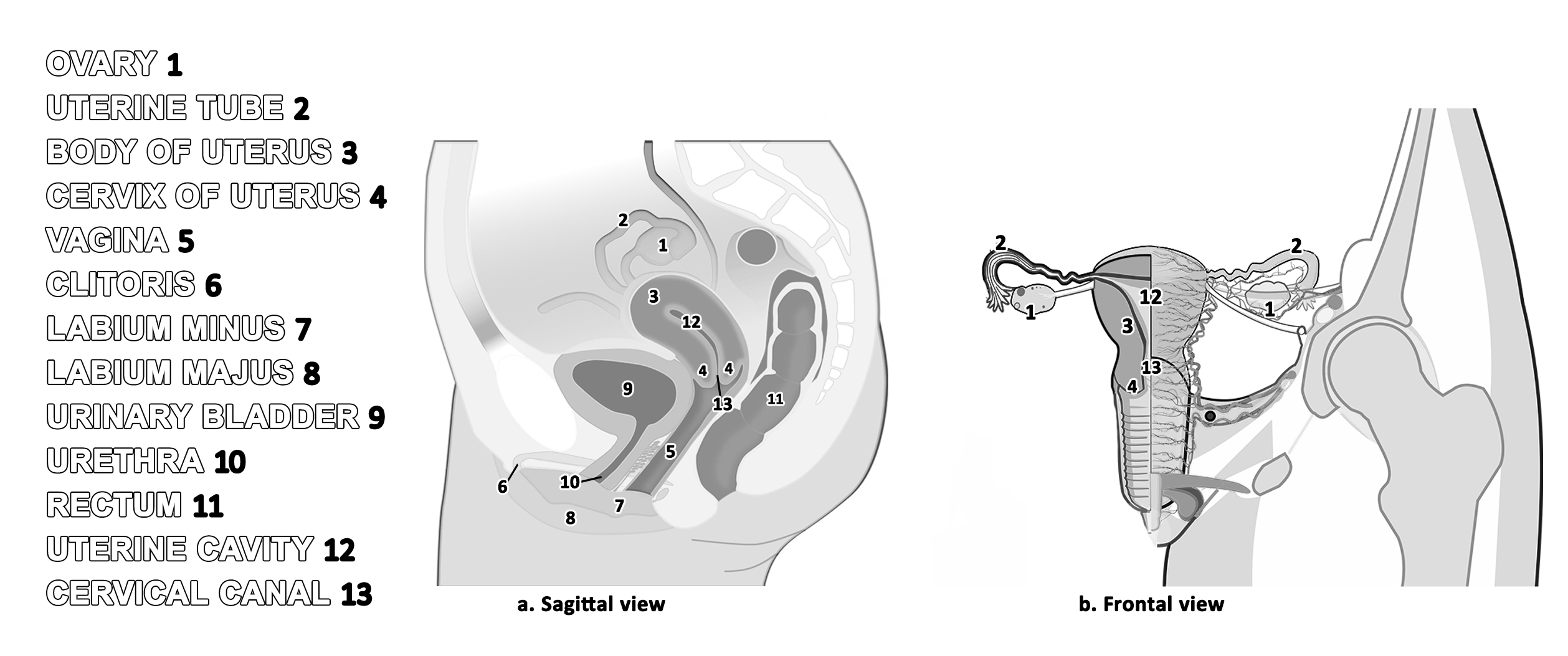
Note: You are not expected to know every term listed on this review page. Review Figure 10.9 to study for the lab practical exam.
Review Activity 10 .4
Help yourself visualize the different body organs in respect to other structures in the system. Using crayons, colored pencils, and/or markers, fill in the name of each organ with a specific color and using that same color fill in the corresponding organ. Use different colors for different organs/structures.
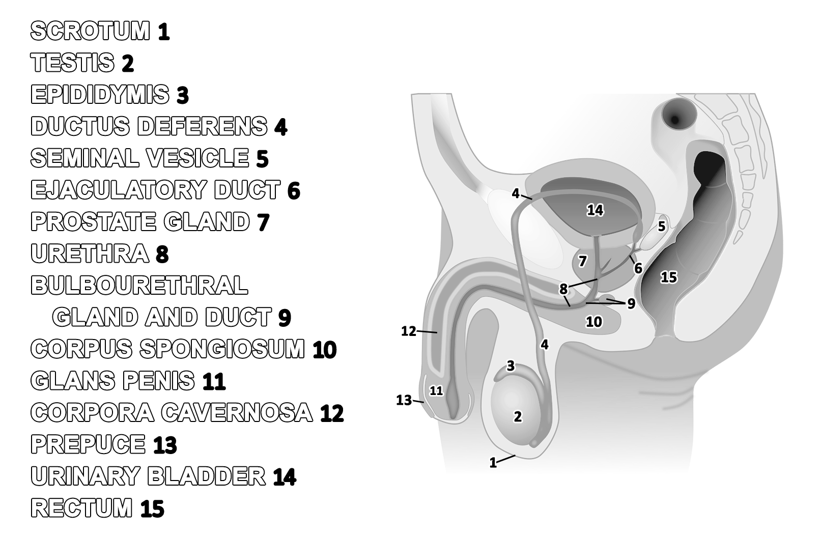
Note: You are not expected to know every term listed on this review page. Review Figure 10.8 to study for the lab practical exam.

_(cropped).png)