
12 Gas Exchange and Respiratory System
Following this week’s lab, students will be able to:
- Identify structures and describe the function of the mammalian respiratory system
- List at least two similarities and two differences each between the respiratory systems of
- fish, insects, and mammals
- Describe the pathway air or water takes through the above animal respiratory systems and where gas exchange occurs
- Define at least four different human lung volumes and explain their significance in a health context
- Demonstrate proper dissection technique and respect for animal specimens and dissection tools
 Contribution Points:
Consult with your TA to receive a stamp at the end of your lab period.
I have completed the necessary tasks required during this week’s lab to earn Contribution Points. I am aware that I may have point(s) deducted from my Contribution Points if my workspace is not appropriately clean at the conclusion of lab.
Your TA will check that you have attempted to complete the dissections associated with this week’s lab activity.
Resources
- Dissection Interchapter 6 “Dissection Techniques and Terminology” (pp. 137–140)
- Fetal pig dissection tutorial video (tutorial video on Canvas)
- Fetal pig dissection guide, Smith and Schenk 2011 (provided in binders for use in lab)
- Biological Science, Freeman et al. 2024 (8th edition)
- Respiratory System (42.1–42.4)
- Internet and Canvas resources
![]() This icon represents a question meant to test your understanding. Check the box once you answer it. Answering these questions in the space provided as you go through the lab will help you better understand the topic and study more effectively. Use your text or e-book, pig dissection guide, and the internet to help you.
This icon represents a question meant to test your understanding. Check the box once you answer it. Answering these questions in the space provided as you go through the lab will help you better understand the topic and study more effectively. Use your text or e-book, pig dissection guide, and the internet to help you.
Part 1: Human Lung Volumes
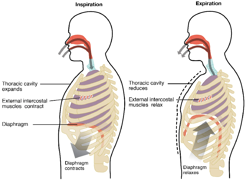
Ever wonder how much air your lungs can hold? When you inhale and exhale, you use muscles to expand your rib cage to let air in and compress your rib cage to push air out. This is a form of ventilation, which is the movement of air or water past a respiratory surface so that gas exchange can occur between the external environment and body tissues. In this case, the respiratory surface is in the lungs, but in other animals it can be a part of gills or tracheae. The diagram below exhibits what happens to your lungs when you inhale (inspiration) and exhale (exhalation). In lab today we will explore what is happening in your lungs during these actions and how efficient your lungs are during normal breathing to better understand how lungs function.
There are several lung volume measurements that are important for assessing an individual’s health and understanding how human lungs function. Table 12.1 shows values for human respiratory volumes for the typical adult. Using this table and information from the video shown in class, work with your group mates to define the following:
Tidal Volume:
Inspiratory reserve volume:
Expiratory reserve volume:
Vital Capacity:
|
Volume |
Value (liters) |
Value (liters) |
|---|---|---|
| Inspiratory reserve volume | 3.1 | 1.9 |
| Tidal volume | 0.5 | 0.5 |
| Expiratory reserve volume | 1.2 | 0.7 |
| Vital capacity | 5.8 | 4.2 |
- The diagram below is an unlabeled drawing depicting the above ventilation volumes. Fill out the blanks below with the name of each segment.
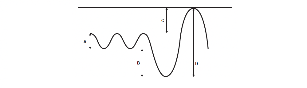
A:
B:
C:
D:
Part 2: Respiratory System Dissections and Stations
During this portion of the lab, fill in Table 12.2 to help organize your knowledge about the respiratory structures of mammals, fish, and insects. You will need the table to complete the post-lab worksheet.
-
Table 12.2. Characteristics of the respiratory system of mammals, fish, and insects. Characteristics Mammals Fish Insects Structures and actions involved in ventilation Respiratory medium (air or water) Structures and actions involved with the respiratory surface Delivery to cell
Fetal Pig Respiratory System Dissection
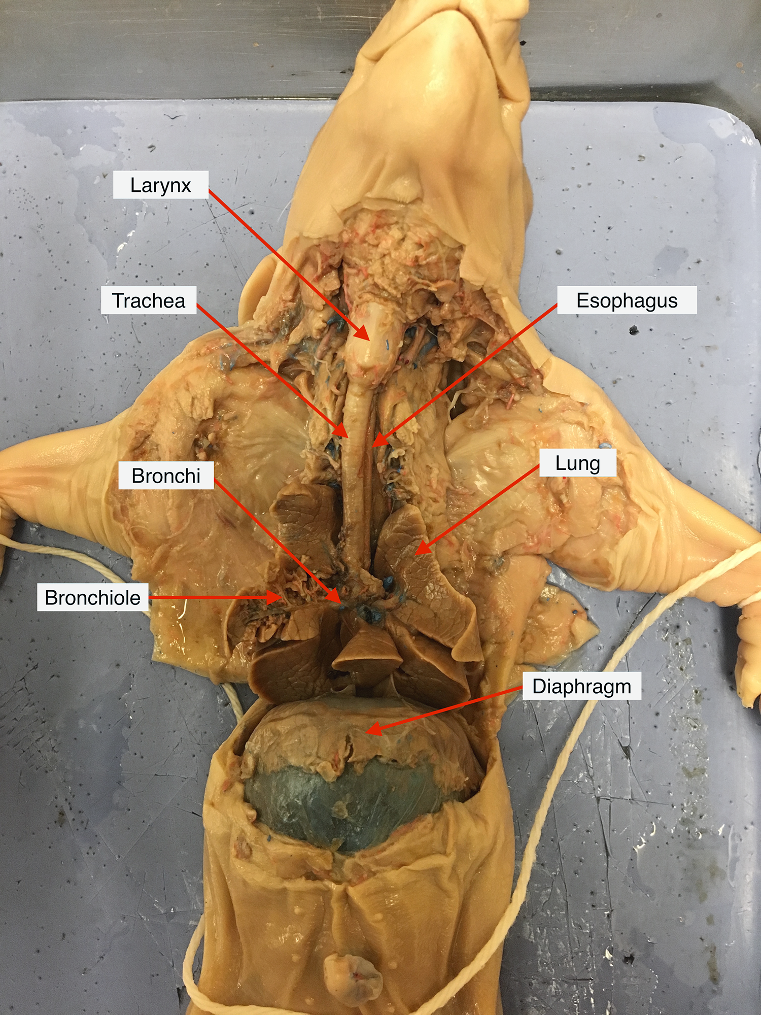
If you haven’t done so already, open your pig’s rib cage to expose its lungs and diaphragm. Remove any extra tissue around the neck, including the thyroid and thymus if you have not removed them already, to expose the trachea and larynx. Identify the organs in the figure below in your pig.
As mammals, we breathe through our nose and mouth and ingest food and water through our mouth. Both the nose and mouth empty into a shared passageway, called the pharynx. The pharynx then leads to both the esophagus and the larynx. We learned about the route of food and water through the esophagus in Laboratory 10. Air goes through the larynx to the trachea and eventually reaches the lungs. It is vitally important that the food we eat does not end up in our lungs! Figure 12.3 (below) shows where the epiglottis is located within the human pharynx.
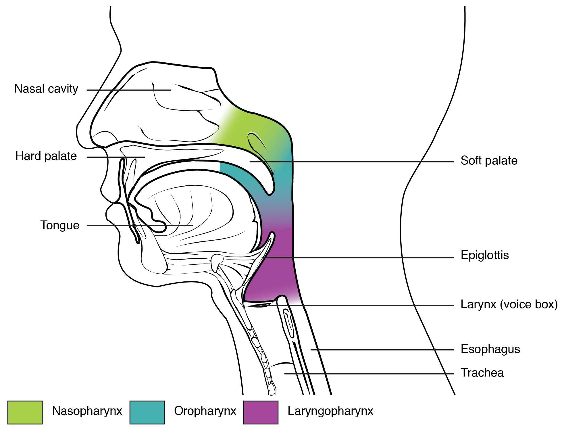
![]() Trace the passage of food through the pharynx into the esophagus on the illustration above.
Trace the passage of food through the pharynx into the esophagus on the illustration above.
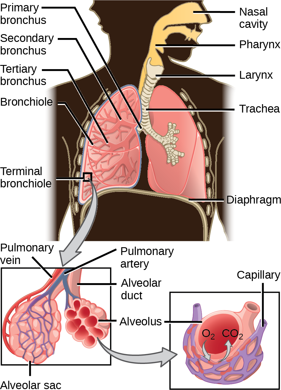
Once air enters the body through the mouth and trachea, it moves from the respiratory tree to the alveoli as described in Figure 12.4 below.
You can’t see some of these structures with the naked eye, so we have provided cross sectional slides of mammal lungs to show you the microscopic structures. Observe this slide at the lab station and sketch what you see below.

Insect Tracheal Respiratory System
Now that you have explored the mammalian respiratory system, it is time to observe the respiratory systems of different organisms. They are all quite different! Go to the insect station in the lab and check out the live hornworms. Look on the side of the hornworm to find the spiracles. They are found on the side of the body, one pair in each abdominal segment. At this station there is also a laptop computer with a video of a giant katydid breathing! You can also find the video linked on Canvas.
![]() What is the function of the spiracles in insects?
What is the function of the spiracles in insects?
![]() Do insects have lungs like mammals? If not, what do they have instead?
Do insects have lungs like mammals? If not, what do they have instead?
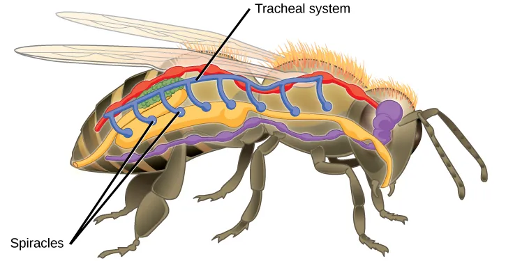
Next, look at the demonstration dissection of a cockroach under the dissecting scope. The shiny mass of tubes you see in the abdomen are called tracheoles (use the photo posted at the station to help you distinguish the tracheoles from the insect digestive system). You may notice air sacs attached to the tracheoles. These are expansions of the tracheoles that are especially important in larger insects to help push air throughout the body. Use Figure 12.5 to identify the structures in a bee.
Fish Respiratory System
Go to the fish station to observe the photos, illustrations, and videos available. Make sure you can identify the operculum, gill arches, and gills.
![]() What is the function of the operculum in fish? What might happen if the operculum was damaged?
What is the function of the operculum in fish? What might happen if the operculum was damaged?
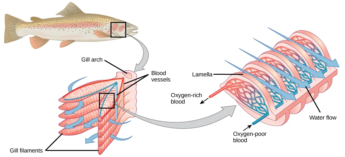
Since fish live underwater, their respiratory system is quite different than ours. Check out Figure 12.6 which depicts the path of water across a fish’s gills.
![]() Refer to the figure above to trace the pathway of water flow over the gills of the fish. How do gills work? What happens to the water as it enters the gills? Where does the water go after it leaves the gills?
Refer to the figure above to trace the pathway of water flow over the gills of the fish. How do gills work? What happens to the water as it enters the gills? Where does the water go after it leaves the gills?
![]() Look at the gill arch slide under the compound microscope. Sketch what you see below.
Look at the gill arch slide under the compound microscope. Sketch what you see below.

![]() Based on your observations, what structural features in the gills increase surface area for gas exchange?
Based on your observations, what structural features in the gills increase surface area for gas exchange?
![]() Why do you think gills stop functioning when a fish is taken out of the water?
Why do you think gills stop functioning when a fish is taken out of the water?
Study Material
The following material will not be collected or graded but is provided to further test your understanding of the material in the lab. These will be valuable assets for quiz and practical studying.
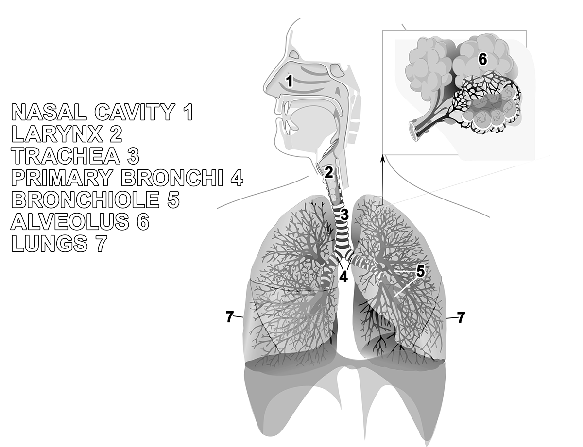
Review Activity 12 .1
Help yourself visualize the external parts of the mammalian respiratory system. Using crayons, colored pencils, and/or markers, fill in the name of each structure with a specific color and using that same color fill in the corresponding structure. Use different colors for different parts.
