
14 Muscular and Skeletal Systems
Following this week’s lab, student’s will be able to:
- Identify the structures and describe the function of the mammalian muscular system
- Identify the structures and describe the function of the mammalian skeletal system
- Describe similarities and differences between the skeletal systems of different vertebrates based on their locomotory behavior
- Describe similarities and differences between the hydrostatic, exo-, and endo-skeletons
- Use PowerLab and Chart software to measure muscle summation, tetany, and recruitment and describe what those terms mean in a physiological context
Contribution Points:
 Consult with your TA to receive a stamp at the end of your lab period.
I have completed the necessary tasks required during this week’s lab to earn Contribution Points. I am aware that I may have point(s) deducted from my Contribution Points if my workspace is not appropriately clean at the conclusion of lab.
Resources
- Fetal pig dissection guide, Smith and Schenk 2011 (provided in binders for use in lab)
- Biological Science, Freeman et al. 2024 (8th edition)
- Muscular and Skeletal System (Chapter 45)
- Canvas resources
![]() This icon represents a question meant to test your understanding. Answering these questions in the space provided as you go through the lab will help you better understand the topic and study more effectively. Use your text or e-book, pig dissection guide, and the internet to help you.
This icon represents a question meant to test your understanding. Answering these questions in the space provided as you go through the lab will help you better understand the topic and study more effectively. Use your text or e-book, pig dissection guide, and the internet to help you.
Printed instructions for the PowerLab Activity can be found at each PowerLab station in the laboratory. They can also be located on Canvas. Students should follow the printed instructions to complete the activity. As the activity is completed in lab, students should answer the related questions in their lab manual.
PART 1: Muscle Physiology PowerLab Activity
Exercise 2: Twitch Response and Recruitment
Complete Exercise 2 and fill in the data table below.
|
Stimulus |
Response (mV.s) |
Stimulus |
Response (mV.s) |
|---|---|---|---|
|
0.0 mA |
|
11.0 mA |
|
|
1.0 mA |
|
12.0 mA |
|
|
2.0 mA |
|
13.0 mA |
|
|
3.0 mA |
|
14.0 mA |
|
|
4.0 mA |
|
15.0 mA |
|
|
5.0 mA |
|
16.0 mA |
|
|
6.0 mA |
|
17.0 mA |
|
|
7.0 mA |
|
18.0 mA |
|
|
8.0 mA |
|
19.0 mA |
|
|
9.0 mA |
|
20.0 mA |
|
|
10.0 mA |
|
|
|
Motor Unit Recruitment Questions.
Muscles are composed of many motor units. Each motor unit consists of a group of muscle cells all controlled by a single motor neuron, and the muscle cells in each motor unit contract in an all-or- none fashion. As additional motor units are stimulated the force of contraction increases (a process known as recruitment).
- What happened to muscle contraction force as you increased the amplitude (in milliamps) of the electrical current stimulus you delivered to the median nerve? Describe what you see in the data you collected.
- Why is there a threshold? Or in other words, a minimum stimulus current below which no contraction occurred? Why was there no contraction below the threshold stimulus current?
- Why does contraction force increase with stimulus amplitude beyond this threshold?
- Why does contraction force reach a maximum, even though stimulus amplitude continues to increase?
Summation and Tetanus
In this exercise, you will keep stimulus amplitude constant at the value that produced a maximal contraction in the preceding experiment, and examine the effects of changing the frequency with which the muscle is stimulated. With increasing frequency of stimulation, the muscle may not relax completely before the next stimulus arrives. As a result, a new contraction begins in a muscle fiber that is already partly contracted.
![]() What effect do you think this might have on the force of contraction of that muscle? State this as a hypothesis:
What effect do you think this might have on the force of contraction of that muscle? State this as a hypothesis:
Follow the printed instructions to complete the activity for Summation and Tetanus. Fill in the data tables below. Then answer the questions on the following page.
Exercise 3: Summation and Tetanus
| Stimulation Frequency (Hz) | Stimulus Interval(s) | Amplitude of First Response (mV.s) | Amplitude of Second Response (mV.s) |
|---|---|---|---|
|
1 |
|
|
|
|
2 |
|
|
|
|
5 |
|
|
|
|
10 |
|
|
|
|
20 |
|
|
|
|
Stimulus Frequency (Hz) |
Stimulus Interval(s) |
Number of Pulses |
Amplitude of Response (mV.s) |
|---|---|---|---|
|
20 |
|
3 |
|
|
20 |
|
4 |
|
Temporal Summation and Tetany. Answer each of the questions (A through D) below.
- When you stimulated your subject’s muscles with bursts of 4 stimuli at different frequencies, you should have seen results similar to those pictured in figure included in Step 8 above. What happened to the force of contraction as stimulation frequency increased? Describe the collected data.
- Was there a frequency at which you saw a marked increase in muscle force from the peak force seen in the first experiment exploring recruitment? If so, what was the threshold frequency for your test subject?
- What chemical process within your muscle cells could account for this increase of maximum muscle force at tetany? What role does Calcium play?
- How do you know that the results of this experiment do not just represent recruitment of additional motor units?
PART 2: Muscle and Skeletal System Stations
Station 1: Microscopic Anatomy of the Neuromuscular Junction and Muscle Tissue
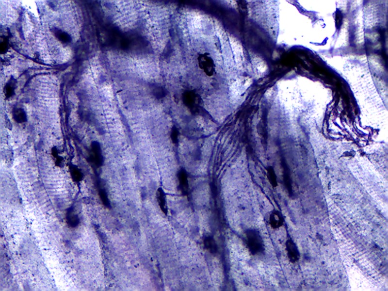
![]() Look at the prepared slide of the neuromuscular junction under a compound microscope. Identify muscle cells, axons of motor neurons, and the synapses between motor neurons and muscle cells, called neuromuscular junctions and label them on Figure 14.1 below.
Look at the prepared slide of the neuromuscular junction under a compound microscope. Identify muscle cells, axons of motor neurons, and the synapses between motor neurons and muscle cells, called neuromuscular junctions and label them on Figure 14.1 below.
![]() What is a neuromuscular junction?
What is a neuromuscular junction?
![]() How many neuromuscular junctions are there in each muscle cell?
How many neuromuscular junctions are there in each muscle cell?
![]() With how many muscle cells does a single neuron synapse?
With how many muscle cells does a single neuron synapse?
![]() Are muscle cells controlled individually, or in groups?
Are muscle cells controlled individually, or in groups?
Look at the slides available of the three different muscle types: smooth, cardiac, and skeletal. Sketch what you see in Table 14.3 and describe the characteristics of each type. Think about how the structure is related to the function of each muscle type.
| Muscle Cell Type | Sketch | Function and Characteristics |
|---|---|---|
|
Smooth |
|
|
|
Cardiac |
|
|
|
Skeletal |
|
|
Station 2: Exoskeletons, Endoskeletons, and Hydrostatic Skeletons
There are many different types of skeletons in the animal kingdom. First check out the exoskeleton mini-station that includes the crayfish and lubber grasshopper.
![]() What is an exoskeleton, and how can you tell that these animals have exoskeletons?
What is an exoskeleton, and how can you tell that these animals have exoskeletons?
![]() What material makes up the exoskeleton of insects? How is it different from bone?
What material makes up the exoskeleton of insects? How is it different from bone?
![]() How do animals with exoskeletons grow in size?
How do animals with exoskeletons grow in size?
Compare the exoskeletons to the endoskeletons. Take some time to observe the specimens available and answer the following questions.
![]() What is an endoskeleton?
What is an endoskeleton?
![]() How do animals with endoskeletons differ from those with exoskeletons in terms of development, growth, and ability to support their body weight?
How do animals with endoskeletons differ from those with exoskeletons in terms of development, growth, and ability to support their body weight?
Compare the exoskeletons and endoskeletons to the hydrostatic skeletons. Take some time to observe the specimens available and answer the following questions.
![]() What is a hydrostatic skeleton? How does a hydrostatic skeleton work? What kinds of organisms have a hydrostatic skeleton?
What is a hydrostatic skeleton? How does a hydrostatic skeleton work? What kinds of organisms have a hydrostatic skeleton?
Station 3: Anatomy of Bones
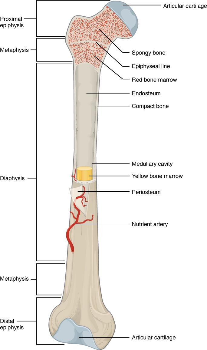
Look at the sectioned femur supplied at station 3 in the lab. Using Figure 14.2 below as a reference, identify the following structures in the sectioned bone: diaphysis, epiphysis, articular cartilage, compact bone, spongy bone, and marrow.
![]() What is a growth plate, and why is it important? In adults, it becomes an epiphyseal line. Why is this?
What is a growth plate, and why is it important? In adults, it becomes an epiphyseal line. Why is this?
![]() Where would you find spongy bone and compact bone? How do they differ in structure and function?
Where would you find spongy bone and compact bone? How do they differ in structure and function?
![]() What are the functions of the red and yellow bone marrow?
What are the functions of the red and yellow bone marrow?
![]() On display at this station are a variety of different kinds of bones from various animals. Make a list of the bones you can identify at this station below.
On display at this station are a variety of different kinds of bones from various animals. Make a list of the bones you can identify at this station below.
Station 4: Bone Materials
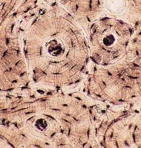
Look at the ground bone prepared slide. Make sure to identify the Haversian (Central) canals, lacunae, and extracellular matrix. Label Figure 14.3 with these structures.
![]() Are bones living or dead in an adult animal? Is your answer consistent with the observation that broken bones can heal?
Are bones living or dead in an adult animal? Is your answer consistent with the observation that broken bones can heal?
![]() What is a Haversian canal, and what would you find in the canal space in a living animal?
What is a Haversian canal, and what would you find in the canal space in a living animal?
Observe the decalcified bones. These vertebrate limb bones have been treated with dilute acid to dissolve the bone mineral hydroxyapatite. Use forceps to remove the decalcified bones to observe them. Put them back when you are done.
![]() What is the material that makes bone hard? What material makes bone somewhat flexible?
What is the material that makes bone hard? What material makes bone somewhat flexible?
![]() How do bone and cartilage differ in both structure and function?
How do bone and cartilage differ in both structure and function?
Station 5: Vertebrate Skeletal Structure and Function
In this station, you will compare the skeletal anatomy of different vertebrates. Be sure to think about how the skeleton of each animal is related to its locomotion and feeding. Some of the skeletons available in lab are: frog, turtle, rat, bat, snake, human, and bird (other skeletons may also be available).
As you observe each skeleton, locate the following bones: vertebrae, skull, sternum, clavicle, humerus, radius, ulna, carpals, metacarpals, ileum, ischium, pubic bone, femur, tibia, fibula, tarsals, and metatarsals.
![]() In the frog, how do the leg bones (tibia/fibula) differ from those in humans and what is the significance of that difference to the frog?
In the frog, how do the leg bones (tibia/fibula) differ from those in humans and what is the significance of that difference to the frog?
![]() How are the forelimbs (front legs/arms) of a frog and bat similar; what features do they share? How are they different and how do the differences relate to the mode of locomotion?
How are the forelimbs (front legs/arms) of a frog and bat similar; what features do they share? How are they different and how do the differences relate to the mode of locomotion?
![]() What adaptations for flight can you see in the skeletons of birds and bats?
What adaptations for flight can you see in the skeletons of birds and bats?
![]() There are strong similarities in the patterning and structure of the skeletons of humans, birds, bats, turtles, frogs, etc. For example, in the forelimb (arm) of these animals, they all have a single humerus, paired radius and ulna, many small carpal bones, and digits, despite differences in locomotion mode and function. What does this suggest about the evolutionary origins of the skeletons of mammals, reptiles, birds, and amphibians?
There are strong similarities in the patterning and structure of the skeletons of humans, birds, bats, turtles, frogs, etc. For example, in the forelimb (arm) of these animals, they all have a single humerus, paired radius and ulna, many small carpal bones, and digits, despite differences in locomotion mode and function. What does this suggest about the evolutionary origins of the skeletons of mammals, reptiles, birds, and amphibians?
Study Material
The following material will not be collected or graded but is provided to further test your understanding of the material in the lab. These will be valuable assets for quiz and practical studying.
Review Activity 14.1
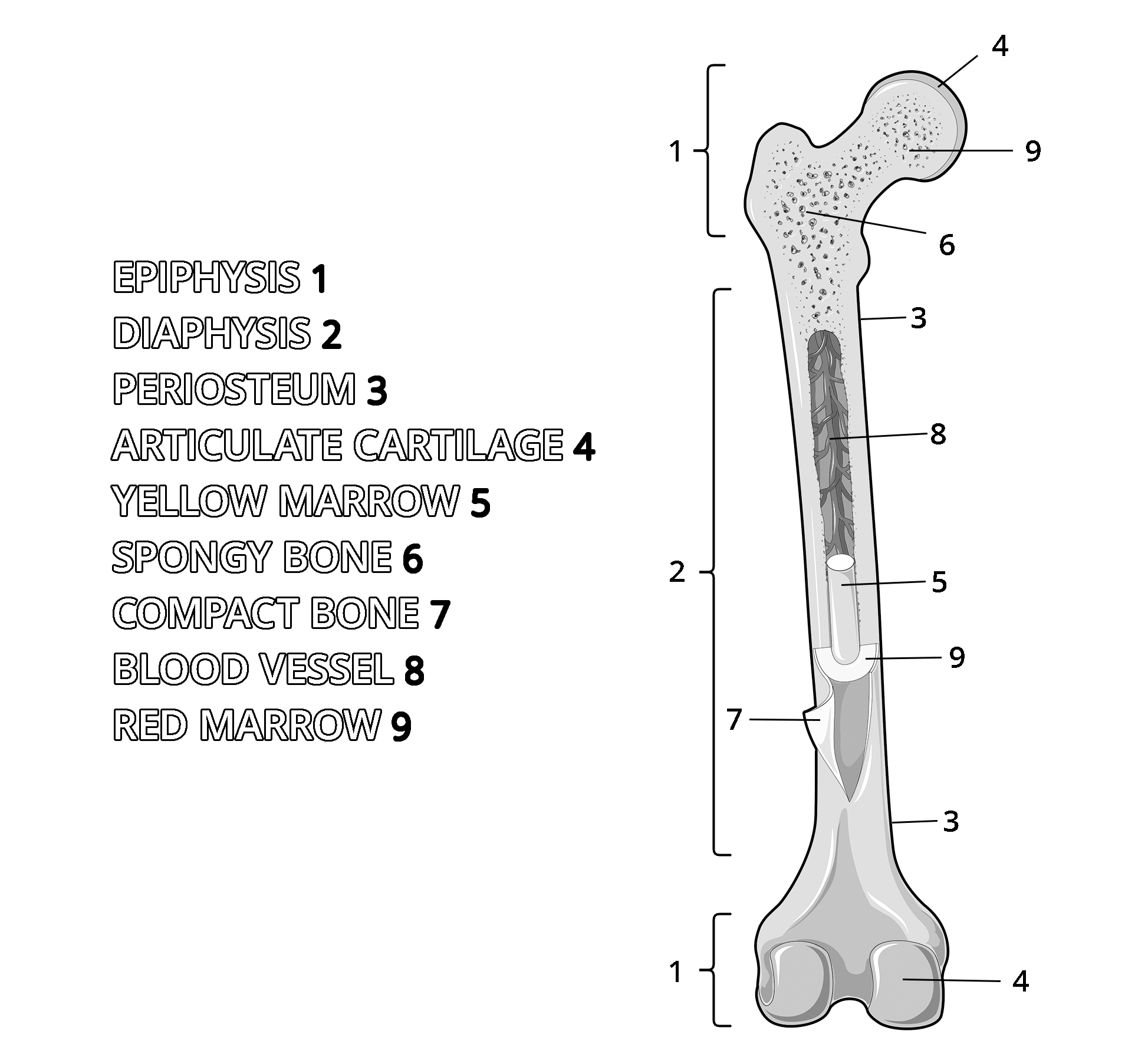
Help yourself visualize the mammalian femur. Using crayons, colored pencils, and/or markers, fill in the name of each structure with a specific color and using that same color fill in the corresponding structure. Use different colors for different parts.
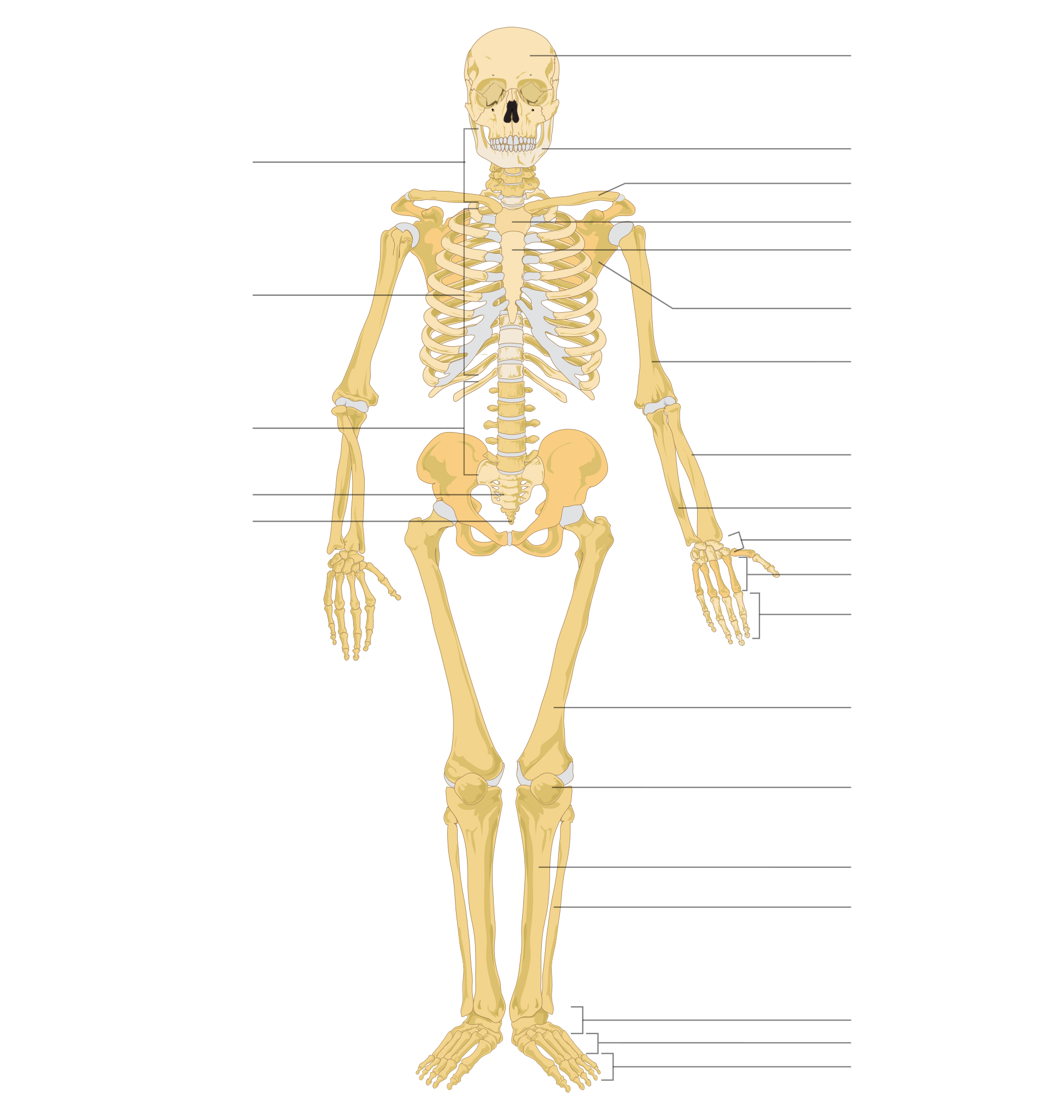
Review Activity 14.2
Test your knowledge of the bones in the human body filling in the missing labels on the diagram of the skeleton below.
