
Gametogenesis, Fertilization, and Implantation
Post Fertilization and Pre-Embryonic Stage
Cleavage and Blastocyst Development
Following fertilization, the zygote and its associated membranes, together referred to as the conceptus, continue to be projected toward the uterus by peristalsis and beating cilia. During its journey to the uterus, the zygote undergoes five or six rapid mitotic cell divisions. The rapid, multiple rounds of cell division are termed cleavage. Although each cleavage results in more cells, it does not increase the total volume of the conceptus. Each daughter cell produced by cleavage is called a blastomere.
Approximately 72 hours after fertilization, a 16-cell conceptus reaches the uterus. The cells that had been loosely grouped are now compacted and look more like a solid mass. The name given to this structure is the morula.
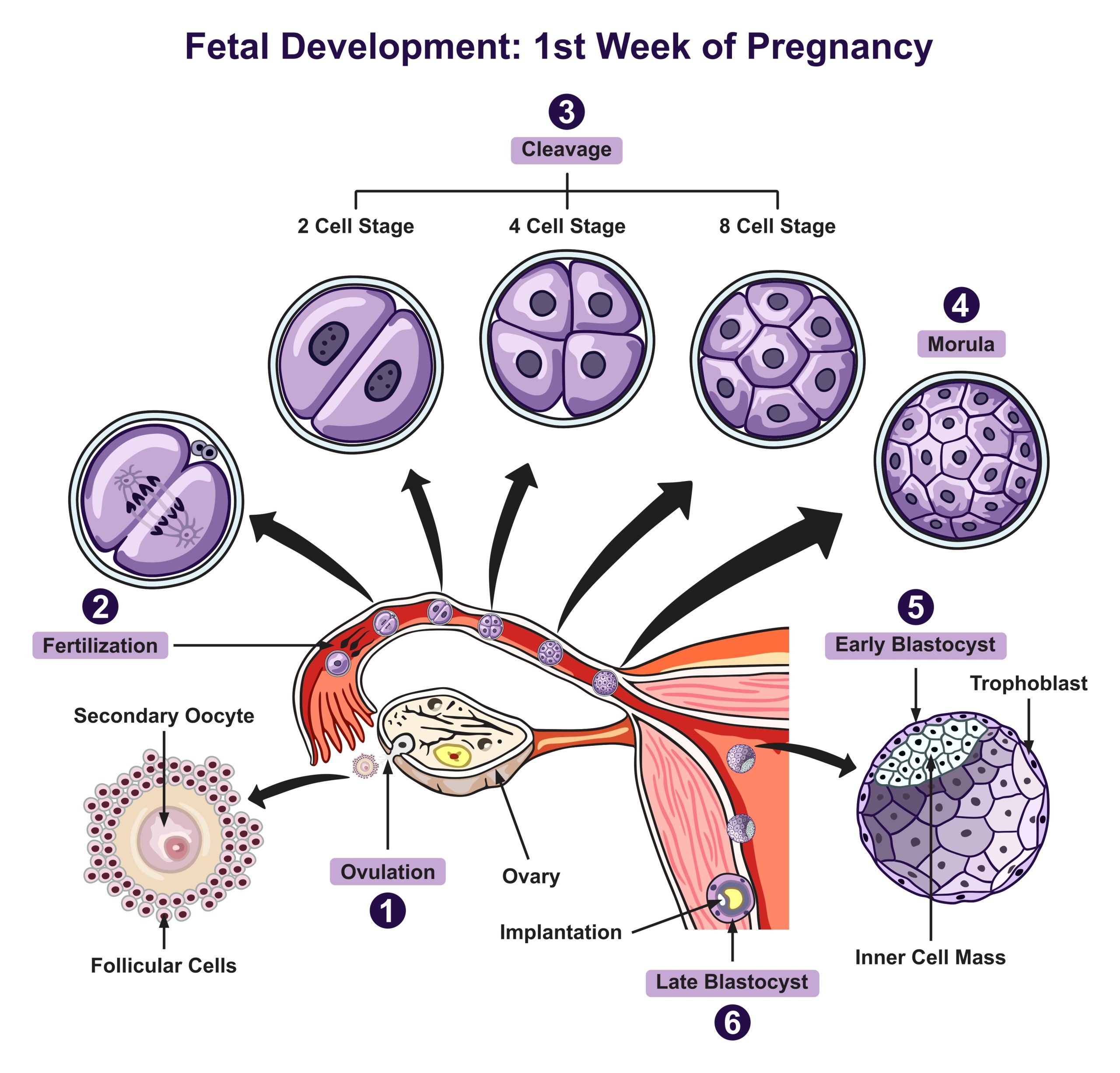
Once inside the uterus, the conceptus floats freely for several more days. It continues to divide, creating a ball of approximately 100 cells and consuming nutritive endometrial secretions called uterine milk while the uterine lining thickens. The ball of the tightly bound cells starts to secrete fluid and form a central fluid-filled cavity. At this developmental stage, approximately 5 days after ovulation, the conceptus is referred to as a blastocyst.
Within the blastocyst, the cells arrange themselves into two layers. A group of cells forms into an inner cell mass which is fated to become the embryo. The cells that form the outer shell, called trophoblasts, will develop into the placenta (the organ of nutrient, waste, and gas exchange between mother and the developing embryo).
As the blastocyst forms, the trophoblast excretes enzymes that begin to degrade the zona pellucida. In a process called “hatching,” the conceptus breaks free of the zona pellucida in preparation for implantation.
Clinical Correlation
Embryonic Stem Cell Research
Up to the 8-cell stage of cleavage, the cells of the conceptus are pluripotent cells, where each cell has the potential to differentiate into any cell type in the human body, and each can be independently developed into an identical conceptus. These cells are called embryonic stem cells (ESC) and are widely used in scientific research to better understand early human development, study diseases, and develop potential treatments. ESCs provide a valuable model for studying cell differentiation and developmental processes; however, research involving ESCs has been a topic of ethical debate because their extraction usually involves the destruction of human embryos.
Identical Twins
Identical twins are the result of a single fertilized egg (oocyte) splitting into two embryos. This splitting typically occurs shortly after fertilization, but it can happen at various stages of development, including before the 16-cell stage.
When the single fertilized egg splits into two embryos, these twins share the same genetic material, making them genetically identical. Because they come from the same fertilized egg, they are of the same sex and usually share a strong resemblance in terms of physical appearance. Identical twins are sometimes called “monozygotic twins” to highlight their single-egg origin.
Take Home Message
- The process by which the cells go into mitotic division after fertilization is known as cleavage.
- The solid mass at 16-cell conceptus is called morula.
- The blastocyst is the fluid-filled space within the conceptus developed approximately 5 days after ovulation.
Image Sources
- Figure 1. “Fetal development first week of pregnancy ” by udaix was licensed through Adobe Stock for educational use.
