
The Female Reproductive System
Female External Genitalia
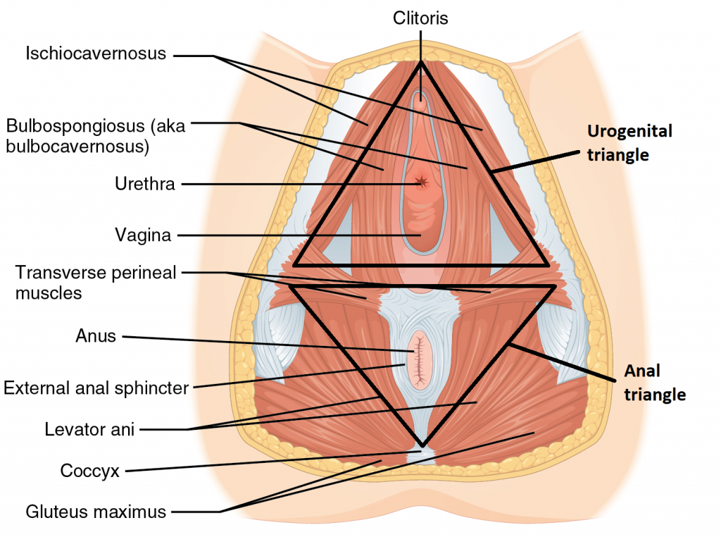
External genitalia are located in the perineal region. The perineum is the diamond-shaped space between the bony landmark, pubic symphysis (anteriorly), the coccyx (posteriorly), and the ischial tuberosities (laterally). Lying just inferior to the pelvic diaphragm (levator ani and coccygeus) until it reaches the skin. It is divided transversely into triangles, the anterior “urogenital triangle,” where the external genitals, the urethral and vaginal orifices in females, are located, and the posterior, “anal triangle,” which contains the anus and external anal sphincter in both sexes (Figure 1).
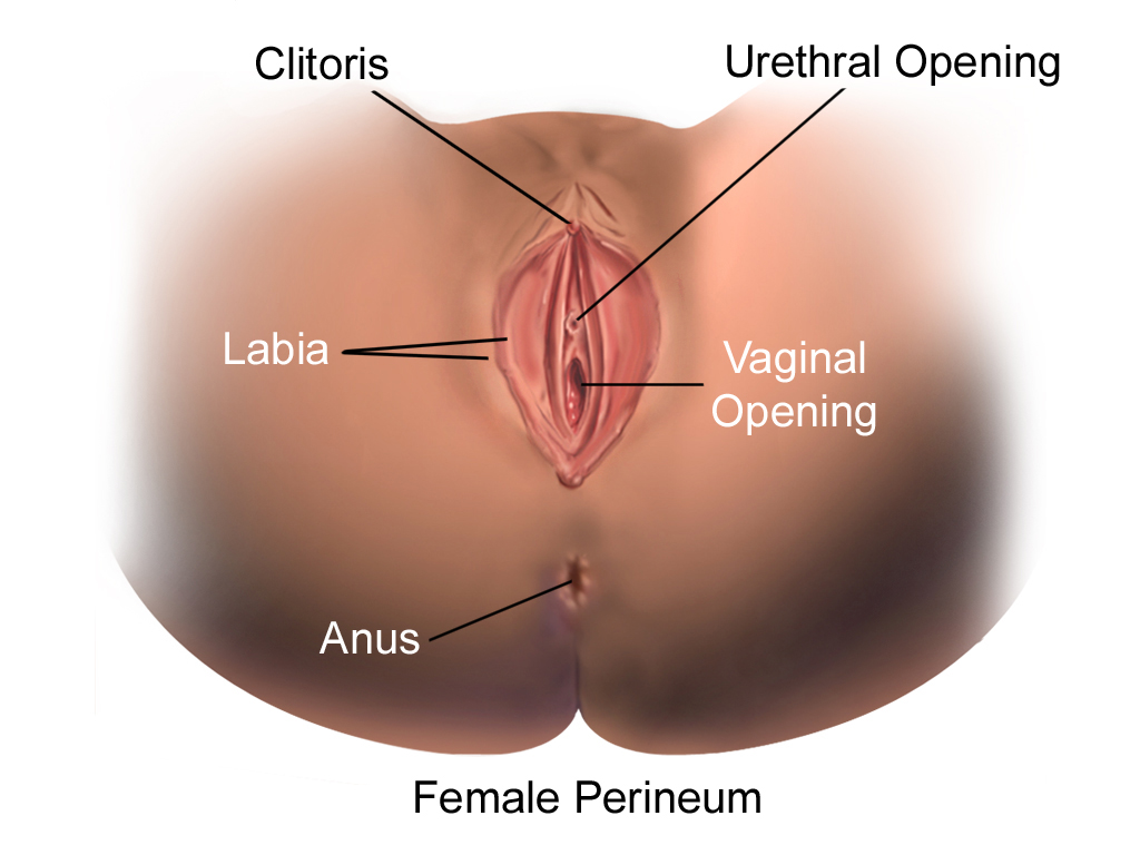
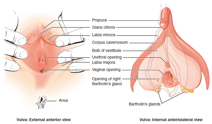
The external female reproductive structures are referred to collectively as the vulva (Figure 2 and 3). The mons pubis is a pad of fat that is located at the anterior, over the pubic bone. After puberty, it becomes covered in pubic hair. The labia majora (labia = “lips”; majora = “larger”) are folds of hair-covered skin that begin just posterior to the mons pubis. The labia majora possess sebaceous and sweat glands and are homologous to the scrotum of the male.
The thinner and more pigmented labia minora (labia = “lips”; minora = “smaller”) extends medial to the labia majora. Although they naturally vary in shape and size from woman to woman, labia minora are devoid of hair and are highly vascularized with numerous melanocytes. The space between the labia minora is the vestibule. Within the vestibule reside the urethral opening, the vaginal orifice, and a pair of greater vestibular glands (glands of Bartholin). The vestibular glands secrete liquid which keeps the vestibular area moist and lubricates the area during sexual intercourse.
The superior, anterior portions of the labia minora come together and form the prepuce, a hoodlike covering over the clitoris. The clitoris is a small (< 2 cm), erectile body, located superior to the urethral opening and homologous to the penis of the male. It consists of two small erectile bodies called the corpora cavernosa form the body of the clitoris and the glands capping the body of the clitoris. The clitoris has abundant nerves that make it important in sexual sensation and orgasm.
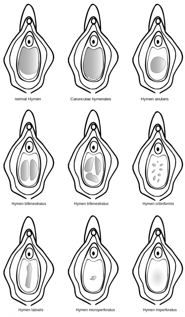
The hymen is a thin perforated membrane that covers the entrance to the vagina. An intact hymen cannot be used as an indication of “virginity”. The hymen can be ruptured with strenuous physical exercise, penile–vaginal intercourse, and childbirth. The hymen is only a partial membrane, as menstrual fluid and other secretions must be able to exit the body. Figure 4 shows different shapes the membrane can take.
Clinical Correlation
Imperforated hymen (Figure 4)
Failure of the hymen to perforate leads to fluid and menstrual blood accumulation and bulge of the membrane. The condition is usually associated with pubic pain at the puberty with the first mensural cycle. Surgical opening and perforation of the membrane can relieve this condition.
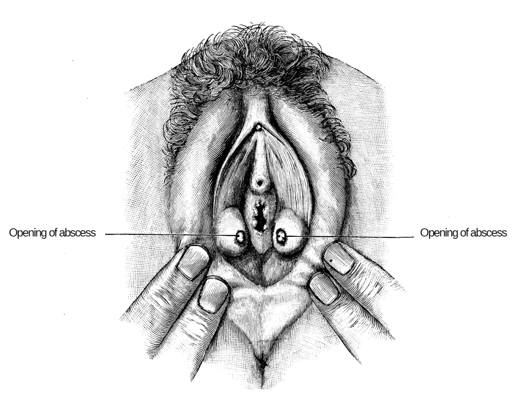
Bartholin’s cyst/abscess (Figure 5)
A Bartholin’s cyst or abscess is a common condition. The openings of Bartholin glands become obstructed, causing fluid to back up and glands became swollen (Bartholin’s cyst). If the fluid becomes infected, that leads to the collection of pus surrounded by inflamed tissue, and an abscess develops. Swollen glands may be painless or, if infected, may became tender with a painful lump. Treatment of a Bartholin’s cyst ranges from good personal hygiene to surgical drainage and antibiotics for infected cysts.
Episiotomy

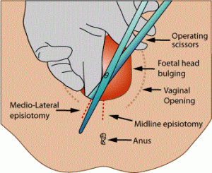
During vaginal childbirth, a significant stretching of the vaginal canal and the perineum occurs, and a potential natural tear of the perineum may happen. Episiotomy, an incision made in the perineum between the vaginal opening and the anus, could be made during childbirth to prevent perineal tearing (Figures 6 and 7). Two types of episiotomy incisions could be done:
- A midline (median) incision, a vertical cut. This type of incision is intended to be easily repairable but carries a higher risk of extending into the anal area and potentially resulting in fecal incontinence.
- A mediolateral incision, an angled cut. While this incision tends to be more painful and somewhat more challenging to repair, it offers greater protection against an extended tear involving the anal area.
Episiotomy was previously done as a routine part of vaginal birth; however, recently it is only done as needed.
Take Home Message
- Female external genitalia are collectively called the vulva. They consist of the mons pubis, labia majora and minora, vestibule, and its component.
- Episiotomy is a planned surgical incision to widen the perineal during vaginal delivery.
- Bartholin glands’ secretions moisten the vestibular area and help lubrication at sexual intercourse.
Image Sources
- Figure 1. “Labeled muscles of the perineum” is adapted from OpenStaxAnatomy & Physiology, licensed CC BY 4.0. Access for free at OpenStax College online.
- Figure 2. “Female perineum” is from Bruce Blaus via Wikimedia Commons, licensed CC BY SA 4.0.
- Figure 3. “The Vulva The external female genitalia” is from OpenStaxAnatomy & Physiology, licensed CC BY 4.0. Access for free at OpenStax College online
- Figure 4. “Hymen membrane placement” is from FollowTheMedia via Wikimedia Commons, licensed CC BY SA 3.0.
- Figure 5. “Batholin’s cyst” is from Ernest, Diseases of Women. Public domain.
- Figure 6. “Possible location of episiotomy” is from Bruce Blaus via Wikimedia Commons, licensed CC BY 3.0.
- Figure 7. “Episiotomy incisions” is from Jeremy Kemp via Wikimedia Commons. Public domain.
