
The Female Reproductive System
Mammary Glands
The mammary glands (breasts) are accessory organs of the female reproductive system located in the thoracic region, far from the other female reproductive organs. The function of the breasts is to supply milk, which contains a complex mixture of proteins, fats, and sugar for infant nutrition in a process called lactation.
The main external feature of the breast is the nipple, a cylindrical projection on the center of the breast containing multiple openings from internal secretory ducts. The nipple is surrounded by a pinkish/brownish pigmented ring of skin called areola (Figure 1), whose coloration deepens during pregnancy. The areola is typically circular and can vary in size from 25 to 100 mm in diameter. The areolar region is characterized by small, raised areolar glands that secrete lubricating fluid during lactation to protect the nipple from chafing. When a baby nurses, or draws milk from the breast, the entire areolar region is taken into the mouth.
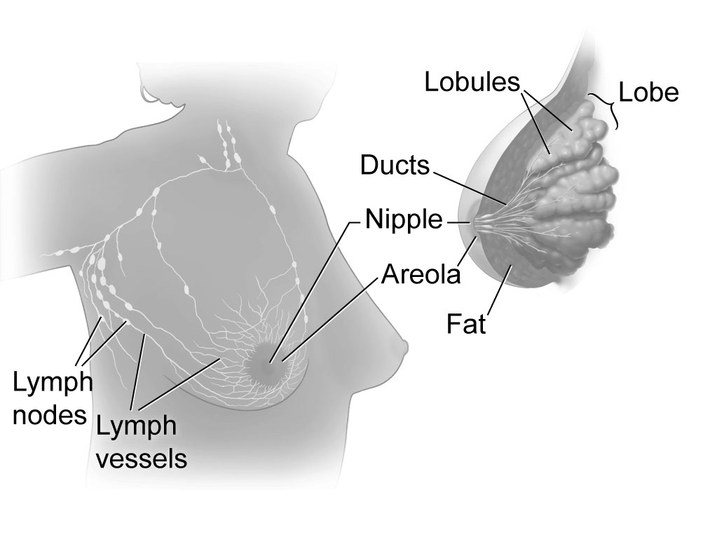
Breast milk is produced by the mammary glands, which are modified sweat glands. The milk itself exits the breast through the nipple via 15 to 20 lactiferous ducts that open on the surface of the nipple. These lactiferous ducts each extend to a lactiferous sinus that connects to a glandular lobe within the breast that contains groups of milk-secreting cells in clusters called alveoli. The clusters can change in size depending on the amount of milk in the alveolar lumen. Once milk is made in the alveoli, stimulated myoepithelial cells that surround the alveoli contract to push the milk to the lactiferous sinuses. From here, the baby can draw milk through the lactiferous ducts by suckling. Prolactin hormone released from the anterior pituitary glands stimulates the process of milk synthesis. Oxytocin hormone secretion by the posterior pituitary, which is stimulated by suckling, leads to alveolus myoepithelial cells contraction and the excretion of milk out of the lactiferous ducts and sinuses.
Internally, the breast is divided into lobes which are further divided into lobules. The lobes are surrounded by fat tissue, which determines the size of the breast. Breast size differs between individuals and does not affect the amount of milk produced. Supporting the breasts are multiple bands of connective tissue called suspensory ligaments that connect the skin of the gland to the overlying fascia of the pectoralis major muscle.
During the normal hormonal fluctuations in the menstrual cycle, breast tissue responds to changing levels of estrogen and progesterone, which can lead to swelling and breast tenderness in some individuals, especially during the secretory phase. If pregnancy occurs, the increase in hormones leads to further development of the mammary tissue and enlargement of the breasts.
Clinical Correlation
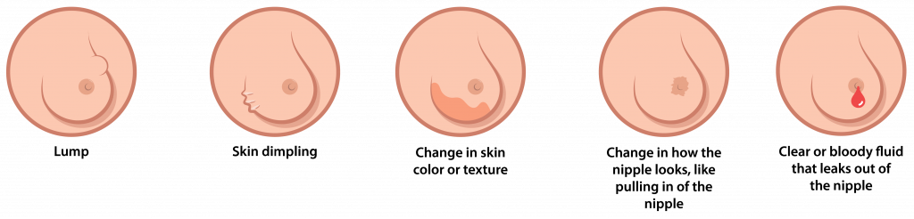
In breast cancer, obstruction of lymphatic drainage leads to swelling and edema in breast tissue while the suspensory ligament remains roped to the skin. This will lead to changes in breast contour and or dimpling appearance, a characteristic feature in breast cancer (Figure 2).
Take Home Message
- Breasts are under the control of estrogen, progesterone, oxytocin, and prolactin hormones.
Image Sources
- Figure 1. “Breast anatomy” is from US National Institutes of Health via Wikimedia Commons. Public domain.
- Figure 2. “External signs of breast cancer” is adapted from Raphseck via Wikimedia Commons, licensed CC BY SA 4.0.
