
Gametogenesis, Fertilization, and Implantation
Gametogenesis
Mitosis and meiosis are the two important cell cycles that aid in the growth and development of the human body and reproduction. Mitosis aids in the growth and replacement of old cells, a process called cytogenesis. Mitosis is a type of asexual reproduction, wherein a single parent diploid cell undergoes division, yielding two diploid daughter cells that are genetically identical to the parent cell.
In contrast, meiosis is a crucial mechanism for generating new cells through sexual reproduction. Meiosis involves two distinct stages, meiosis I and meiosis II, which are essential during gametogenesis to produce four unique haploid daughter cells.
In both cycles, DNA replication occurs first, where the nuclear membrane breaks down and the organism’s DNA condenses to form chromosomes. A key difference between mitosis and meiosis is that during meiosis, when the chromosomes come together, they “cross over,” which allows for the exchange and “mixing” of the DNA coding. After that, the chromosomes separate and migrate to the opposite ends of the cell. Upon the completion of meiosis I and II, the cell divides into 4 distinct, haploid daughter cells, each with its own characteristics.
Gametogenesis is the process of producing gametes, specialized reproductive cells. This process occurs through the cell cycle of meiosis and produces cells with a haploid number of chromosomes. The process of producing gametes in males is called spermatogenesis, and in females, it is called oogenesis. Spermatogenesis leads to the formation of sperm, while oogenesis results in the development of eggs (ova). These processes occur in the testes for males and the ovaries for females, respectively, and are essential for sexual reproduction.
Spermatogenesis
Spermatogenesis occurs in the seminiferous tubules that form the bulk of each testis. The process begins at puberty, after which time sperm are produced constantly throughout the lifespan. One production cycle, from spermatogonia to formed sperm, takes approximately 64 days. A new cycle starts approximately every 16 days, although this timing is not synchronous across the seminiferous tubules. The total number of sperms a man produces slowly declines after age 35, and some studies suggest that smoking can lower sperm counts irrespective of age.
The process of spermatogenesis begins with mitosis of the diploid spermatogonia. Because these cells are diploid (2n), they each have a complete copy of the father’s genetic material, 46 chromosomes. However, mature gametes are haploid (1n), containing 23 chromosomes—meaning that daughter cells of spermatogonia must undergo a second cellular division through the process of meiosis.
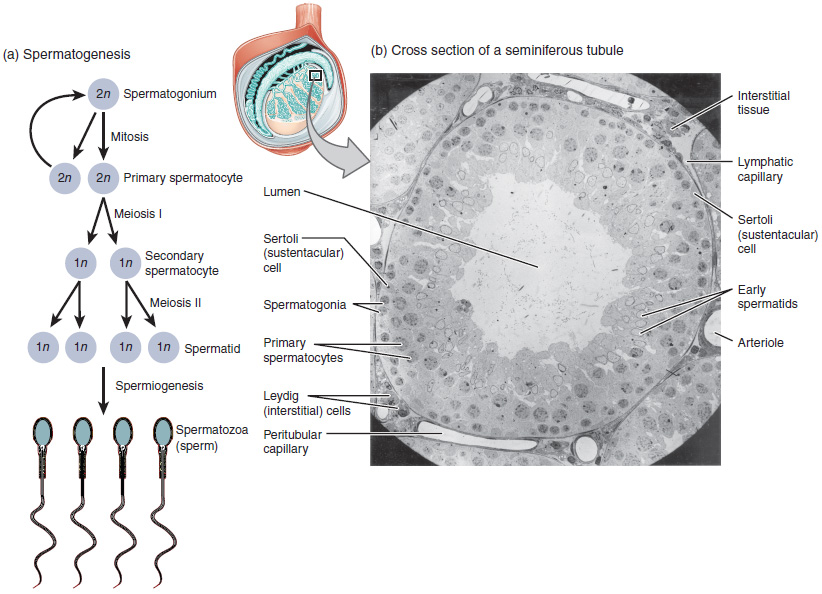
The cycle starts at puberty, where the spermatogonium cell goes through mitosis. Two identical diploid cells result from spermatogonia mitosis. One of these cells remains a spermatogonium, and the other becomes a primary spermatocyte. The next stage is the process of spermatogenesis. As in mitosis, DNA is replicated in a primary spermatocyte before it undergoes a cell division called meiosis I. During meiosis I, each of the 23 pairs of chromosomes separates. This results in two cells, called secondary spermatocytes, each with only half the number of chromosomes. Then, a second round of cell division (meiosis II) occurs in both secondary spermatocytes. During meiosis II, each of the 23 replicated chromosomes divides, similar to what happens during mitosis. Thus, meiosis results in separating the chromosome pairs. This second meiotic division results in four cells with only half the number of chromosomes. Each of these new cells is a spermatid. Although haploid, early spermatids look very similar to cells in the earlier stages of spermatogenesis, with a round shape, central nucleus, and a large amount of cytoplasm. A process called spermiogenesis transforms these early spermatids, reducing the cytoplasm and beginning the formation of the parts of a true sperm. The fifth stage of germ cell formation—spermatozoa or formed sperm—is the end result of this process, which occurs in the portion of the tubule nearest the lumen. Eventually, the sperm are released into the lumen and are moved along a series of ducts in the testis toward a structure called the epididymis for the next step of sperm maturation.
Oogenesis
Oogenesis process begins with the ovarian stem cells, or oogonia. Oogonia are formed during fetal development and divide via mitosis, much like spermatogonia in the testis. Unlike spermatogonia, however, oogonia forms primary oocytes in the fetal ovary prior to birth. These primary oocytes are then arrested in this stage of meiosis I, only to resume it years later, beginning at puberty and continuing until the woman is near menopause (the cessation of a woman’s reproductive functions). The number of primary oocytes present in the ovaries declines from one to two million in an infant, to approximately 400,000 at puberty, to zero by the end of menopause.
The initiation of ovulation—the release of an oocyte from the ovary—marks the transition from puberty into reproductive maturity in females. From then on, throughout the reproductive years, ovulation occurs approximately once every 28 days. Just prior to ovulation, a surge of luteinizing hormone triggers the resumption of meiosis in a primary oocyte. This initiates the transition from primary to secondary oocyte. However, as shown in the figure below, this cell division does not result in two identical cells. Instead, the cytoplasm is divided unequally, and one daughter cell is much larger than the other. This larger cell, the secondary oocyte, eventually leaves the ovary during ovulation. The smaller cell, called the first polar body, may or may not complete meiosis and produce second polar bodies; in either case, it eventually disintegrates. Therefore, although oogenesis produces up to four cells, only one survives.
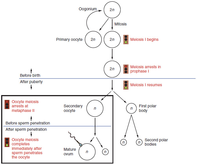
How does the diploid secondary oocyte become an ovum—the haploid female gamete? Meiosis of a secondary oocyte is completed only if a sperm succeeds in penetrating its barriers. Meiosis II then resumes, producing one haploid ovum that, after fertilization by a (haploid) sperm, becomes the first diploid cell of the new offspring (a zygote). Thus, the ovum can be considered a brief, transitional, haploid stage between the diploid oocyte and diploid zygote.
The larger amount of cytoplasm contained in the female gamete is used to supply the developing zygote with nutrients between fertilization and implantation into the uterus. Interestingly, sperm contribute only DNA at fertilization—not cytoplasm. Therefore, the cytoplasm and all of the cytoplasmic organelles in the developing embryo are of maternal origin. This includes mitochondria, which contain their own DNA. Scientific research in the 1980s determined that mitochondrial DNA was maternally inherited, meaning that you can trace your mitochondrial DNA directly to your mother, her mother, and so on back through your female ancestors.
Take Home Message
- Spermatogenesis starts at puberty while oogenesis starts before birth.
- A spermatid is a large cell that loses its cytoplasm and eventually becomes sperm.
- Primary oocytes enter meiosis and stop at the prophase stage of meiosis I. They complete meiosis I at ovulation.
- An LH peak is required for secondary oocyte development.
- Fertilization stimulates the completion of meiosis II and ova development.
