
The Male Reproductive System
Ejaculate
The fluid coming from the three accessory glands combines with sperms from the testes to make semen. When it is released during intercourse, the semen is called ejaculate. The ejaculate is normally about 3-5 ml in volume and contains 200-500 million sperm. Any disturbance of semen quantity or quality will interfere with the capability of fertilization and conception. Evaluation of the semen could be done to detect sperm morphology and motility as a first line for dealing with infertility. An individual can have up to 40% abnormal sperm and still be considered fertile.
Normal Structure of Sperm
Sperm are smaller than most cells in the body; in fact, the volume of a sperm cell is 85,000 times less than that of the female gamete. Approximately 100 to 300 million sperm are produced daily, whereas women typically ovulate only one oocyte per month.
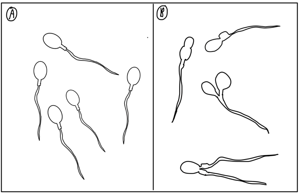
Sperm have a distinctive head, mid-piece, and tail region (Figure 1).
The head of the sperm contains an extremely compact nucleus with a haploid number of chromosomes and very little cytoplasm. These contribute to the overall small size of the sperm (the head is only 5mm long). A structure called the acrosome covers the anterior 2/3 of the nucleus at the head of the sperm cell, the “Acrosomal cap”. This cap is filled with lysosomal and hydrolytic enzymes that participate and initiate acrosomal reactions at the time of fertilization. The acrosomal reaction helps sperm bind and burrow through the Zona Pellucida surrounding the egg and complete the process of fertilization.
The constriction between the head and the mid-piece called the neck.
The mid-piece of the sperm contains a tightly packed mitochondrion that provides the ATP and energy required for movement. The tail, which extends from the mid-piece, consists of a principal flagella structure around 45µm long that is highly mobile, and an end piece, a 5 µm long fibrous sheath of disorganized flagellum structure. Movement of the entire sperm cell occurs through the tail.
Clinical Correlation
Abnormal Semen Disorders
- Aspermia is simply the condition in which an individual cannot produce an ejaculate, while Azoospermia is the condition in which a male makes ejaculate, but it is completely devoid of spermatozoa.
- Oligospermia is when a semen sample has less than 20 million spermatozoa per milliliter of semen.
- Teratospermia is the condition when over half of the sperms present in the ejaculate are of abnormal shape.
- Asthenospermia is when over half of the sperm cells are immobile.
Take Home Message
- Good quantity and quality of sperms are necessary for the process of fertilization and male fertility.
